生理学_循环系统
上半
- Anatomy of the heart
心脏解剖学- location & size
- The heart lies between the two lungs, just above the diaphragm隔膜
- From child to adult, the heart is roughly the same size as the fist
- weight about 300g
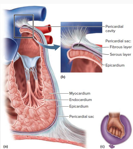
- The pericardium心包 and heart wall
peri- 外围的- 图
- The heart is enclosed in a double-walled sac called the pericardium
- The pericardium protects the heart, and allows it to beat with minimal friction
- The heart wall consists of 3 layers: epicardium (心外膜), myocardium (心肌层) and endocardium (心内膜)
- The myocardium is the thickest layer. It is composed of cardiac muscle, and performs the work of the heart.
- 图
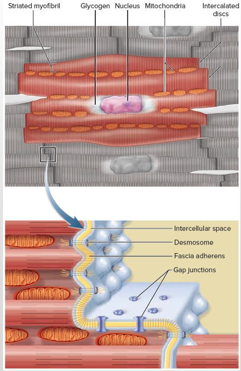
- The cardiac muscle
- The cardiac muscle cells are arranged in spiral bundles
- The plasma membrane at the end of the cardiac cell is folded like the bottom of an egg carton
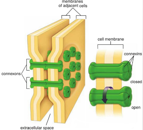
- Gap junctions are important components of the intercalated discs (闰盘)
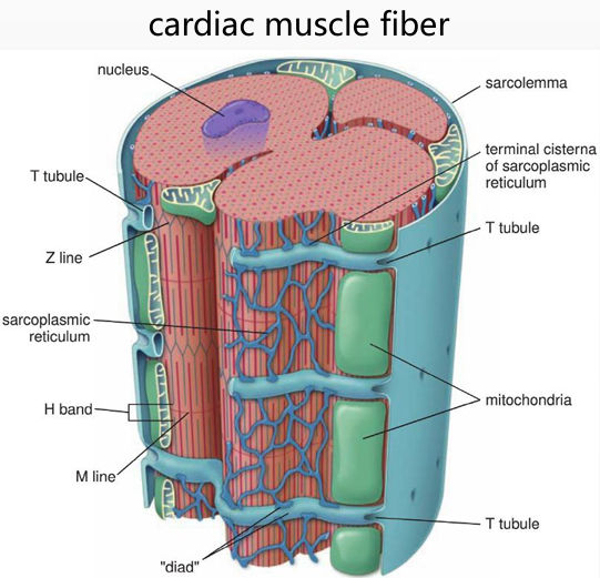
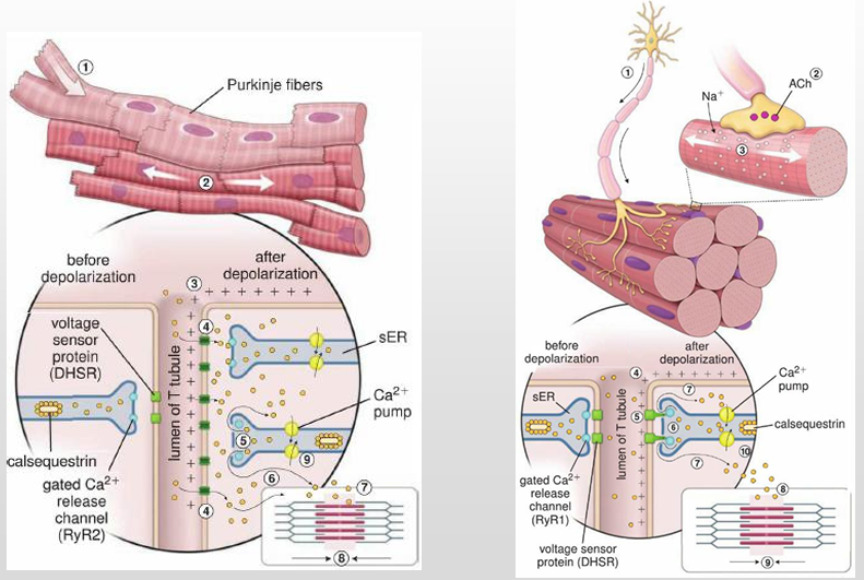
- The sarcoplasmic reticulum is less developed than in skeletal muscle; it forms a diad with the T tubule
- location & size
- RP&AP
- The RP of cardiac muscle
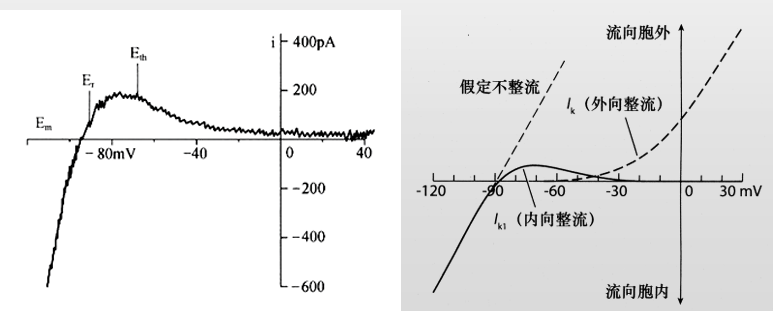
- The resting potential of cardiac working cell =-90mV
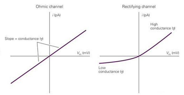
- The inward rectifier K+ channel (内向整流IK1通道) contributes to the resting potential
- rectifier整流通道类似单向门,ohmic欧姆通道两侧都能过
- 让细胞静息电位保持在-90mV
- The AP of cardiac muscle
- 分类
- 根据velocity of depolarization
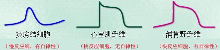
- fast response cell
- rapid depolarization, large amplitude, slow repolarization with a plateau
- rapid depolarization, large amplitude, slow repolarization with a plateau
- slow response cell
- slow depolarization, small amplitude, rapid repolarization without a plateau
- slow depolarization, small amplitude, rapid repolarization without a plateau
- fast response cell
- 根据spontaneous depolarization
- authorhythmic cell
- self-excitation with rhythmical discharge, but can not contract
- working cell
- excited by autorhythmic cell, can contract
- authorhythmic cell
- 根据以上两个分类
- fast-response working cells: atrial心房 muscle and ventricular心室 muscle
- slow-response autorhythmic cells: sinoatrial node窦房结 and atrioventricular node房室结
- fast-response autorhythmic cells: Purkinje fiber, bundle of His, bundle branch
- 根据velocity of depolarization
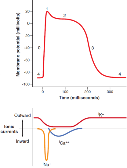
- AP of ventricular muscle
- 图
- 过程
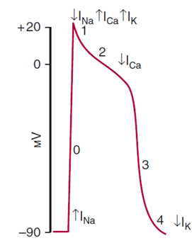
- phase 0: depolarization, INa channels open, 1~2ms
INa导致去极化 - phase 1: initial repolazrization, INa channels close, Ito(transient outward current) channels open, 10ms
K+通道Ito短暂开启复极化 - phase 2: plateau, ICa-L (long-lasting) channels open and counter K+ efflux via IK1 and IK (delayed rectifier K+ current) channels, 100~150ms
Ca2+与两种K+通道导致一段平台期 - phase 3: rapid repolarization, ICa-L channels close, IK1 and IK increase 100~150ms
Ca2+关闭,K+内向整流越来越快地复极化 - phase 4: resting state, Na+-K+ pump, Ca2+ pump and Na+-Ca2+ exchanger together return the membrane potential to -90mV
各种泵恢复静息电位
- phase 0: depolarization, INa channels open, 1~2ms
- 图
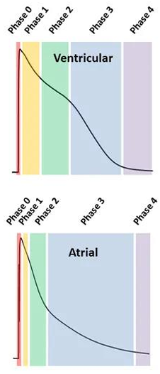
- AP of atrial muscle
- 相比于心室细胞,由于膜上表达通道蛋白不同,心房细胞平台期较短,静息电位为-80mV(Ik1表达较少)
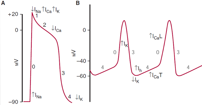
- AP of pacemaker cell
- 与心室细胞相比
- 图
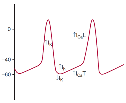
- lower resting potential – lack of Ik1
- lower threshold potential阀电位 and smaller AP amplitude – lack of INa and ICa-L-dependent depolarization
Ca2+导致去极化而不是Na+ - no phase 1 – lack of Ito
- no phase 2 – strong outward Ik can not maintain the balance of the plateau
- 图
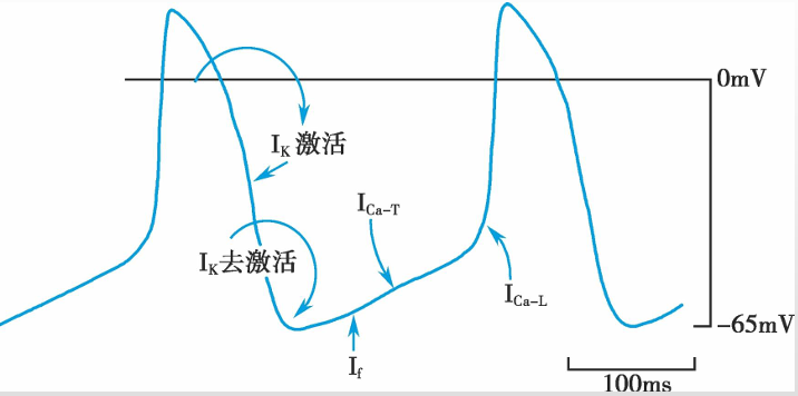
- spontaneous 自主性的原理
- 图
- Ik decline and If (hyperpolarization-activated inward current) activation in phase 4 leads to spontaneous depolarization.
If,funny,在极化(-50mv)时候被激活 - Ik decline is more important.
- When the membrane potential depolarizes to -50mV, ICa-T occurs and then activates ICa-L
- 图
- 与心室细胞相比
- AP of Purkinje fiber
- 快反应自主性
- similar to the one of ventricular muscle
- can depolarize spontaneously due to IK decline and If activation
- If is fully activated at -100mV, and Purkinje fibers repolarize close to this level, thus If is more important
- 快反应自主性
- 分类
- The RP of cardiac muscle
- The electrophysiological properties of cardiac muscle
- excitability
- Factors
- ①The difference between the resting potential / maximal repolarization potential and the threshold potential – small difference means high excitability
静息电位与阀电位差距↓ excitability↑ - ②The status of ion channels that depolarize the membrane potential
- ①The difference between the resting potential / maximal repolarization potential and the threshold potential – small difference means high excitability
- 各个时期
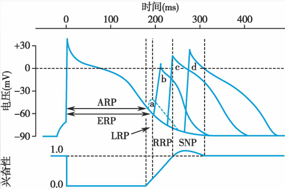
- RP = refractory period
- 绝对不应期ARP
- 有效不应期ERP = ARP + LRP
- 局部反应期LRP
- 相对不应期RRP
- 超常期SNP
- RP = refractory period
- Factors
- conductivity
- 细胞间通过intercalated discs闰盘传递AP
- 过程
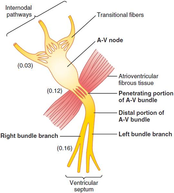
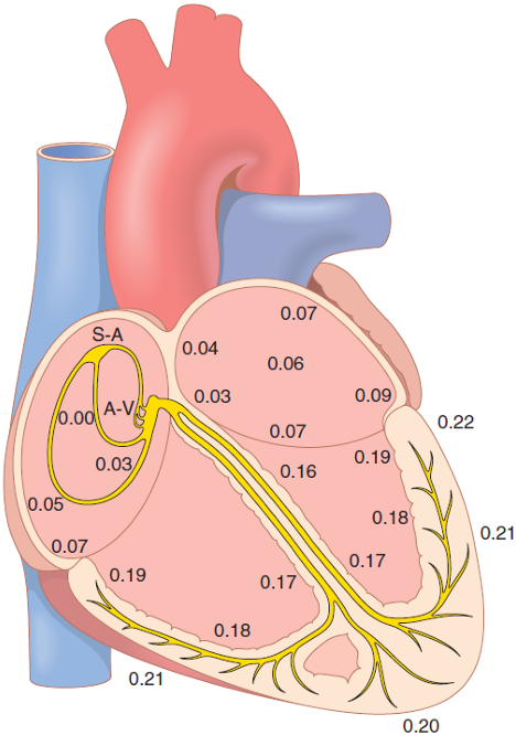
- ①Action potentials originating in the sinus node spread through the entire atrial muscle mass and to the A-V node (房室结)
- ②atrioventricular delay ( 房室延搁) allows time for the atria to empty their blood into the ventricles before ventricular contraction begins
- ③Purkinje fibers lead from the A-V node through the A-V bundle (房室束) into the ventricles. They are very large fibers, and they transmit action potentials at a velocity about 6 times that in the ventricular muscle and 150 times that in some of the A-V nodal fibers
浦肯野纤维连着末端的心室肌肉,传电速度超级快 - 传播时间
- Factors
- ①Structural: fiber diameter, cell differentiation, numbers of gap junctions
- ②Physiological: velocity and amplitude of depolarization in phase 0, excitability of adjacent cells
- 细胞间通过intercalated discs闰盘传递AP
- autorhythmicity
- Discharge rates
- sinoatrial(sinus) node, 70 times/min
- A-V node, 50 times/min
- Purkinje fiber, 25 times/min
- The sinoatrial node controls the beat of the heart by
- ① preoccupation (抢先占领)
- ② overdrive suppression (超速驱动压抑)
- Discharge rates
- excitability
- Myocardial contraction
心肌收缩- 相比于肌肉,心肌没有triad只有diad
- 特点
- ①Simultaneous contraction
- all-or-none contraction.
- The cardiac muscle fibers of atria and ventricles function as two syncytiums (合胞体) due to the intercalated discs.
- ②Dependence of extracellular Ca2+
- The sarcoplasmic reticulum in the cardiac muscle is less developed than in the skeletal muscle, providing only 80~90% Ca2+ for contraction
- ③No tetanus强直收缩
- Prolonged ERP lasts till muscle relaxation begins
有效不应期到肌肉开始放松才结束
- Prolonged ERP lasts till muscle relaxation begins
- ①Simultaneous contraction
- 相比于肌肉,心肌没有triad只有diad
- Electrocardiogram
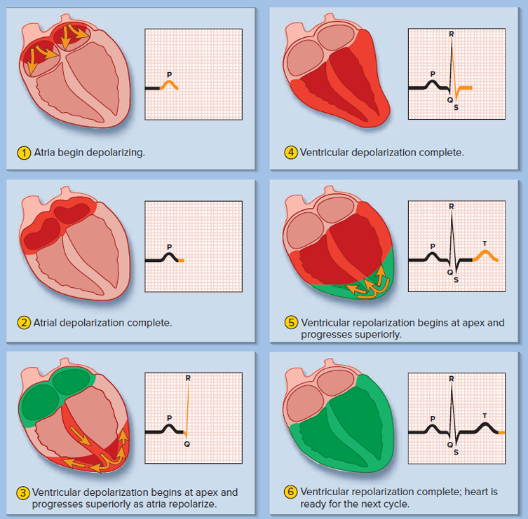
- P-QRS-T, depolarization,repolarization
- P wave – atrial depolarization
- QRS complex – ventricular depolarization
- T wave – ventricular repolarization
- PR interval – atrioventricular delay
- QT interval – ventricular AP/ contraction
- ST segment – plateau of ventricular AP
- P-QRS-T, depolarization,repolarization
- The heart as a pump
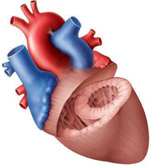
- Internal anatomy of the heart
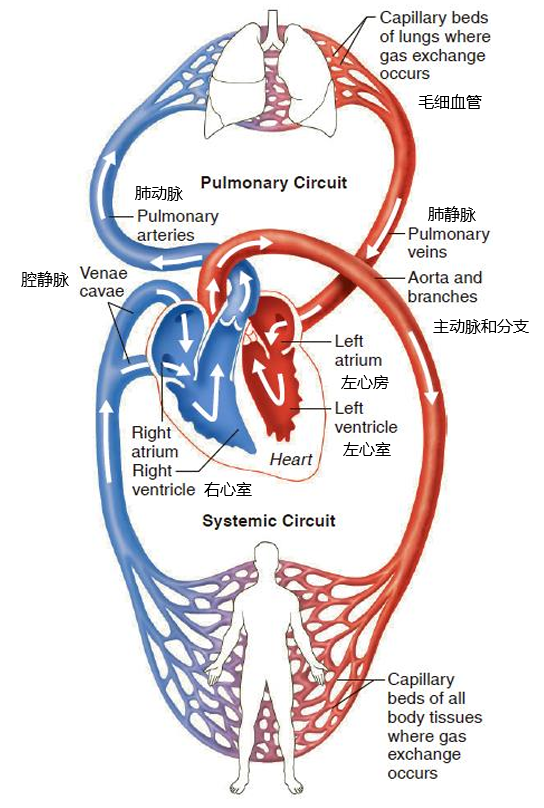
- 右边连着静脉,左边连着动脉,肺动脉里是静脉血,肺静脉里是动脉血
- arteries动脉,veins静脉,pulmonary肺,aorta主动脉,capillary毛细血管,atrium心房,ventricle心室
- 右心室是个月亮,左心室是个圆
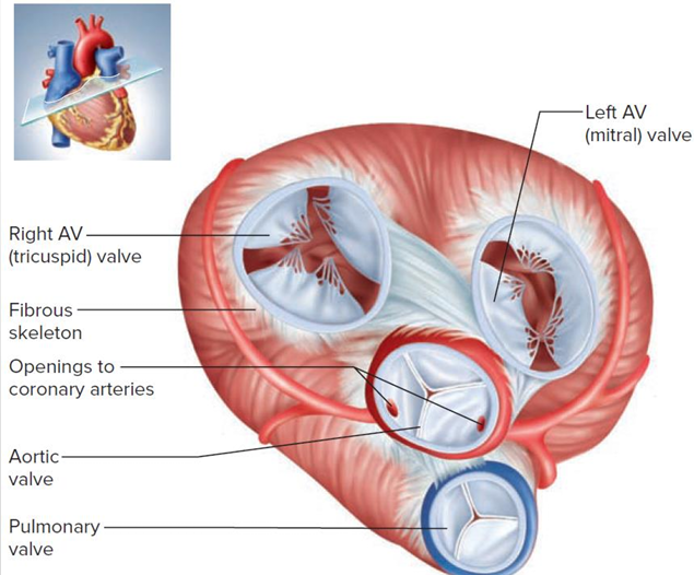
- The atrioventricular valves房室瓣 are anchored to the papillary乳突的 muscles of the ventricle by the chordae tendineae (腱索)
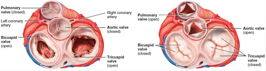
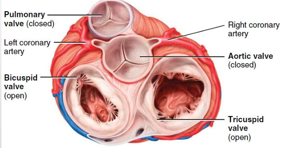
- 左侧semilunar (SL) valves半月瓣 closed, atrioventricular valves open 右侧相反
- 右边的房室瓣atrioventricular valve是三尖瓣tricuspid valve,左边的房室瓣atrioventricular valve是双尖瓣bicuspid valve = mitral valve
- 右边连着静脉,左边连着动脉,肺动脉里是静脉血,肺静脉里是动脉血
- The cardiac cycle
- 右心室压力变化比左心室小
- systole收缩,diastole舒张,心跳变快时候,舒张时间显著减少
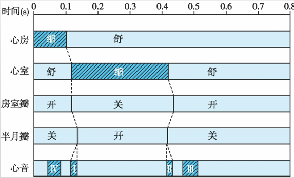
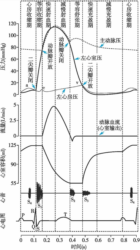
- 看图说话环节,下面蓝色的是ventricular systole,红色的是ventricular diastole,Pao动脉压力,Pv心室压力,PA心房压力
- 心房收缩期atrial systole
- the end of ventricular diastole
- 心房和心室压力缓慢增加
- 心房压力大于心室压力,bicuspid valve打开,1/3的体积流入心室
- P波表示这个时期的开始
- 等容收缩期isovolumetric contraction
- 心室压力>心房压力,bicuspid valve关闭,有一个强劲的声音
- 心室压力<动脉压力,体积不变化没有血液流出
- QRS波表示这个时期的开端
- 快速射血期rapid ejection
- 心室压力>动脉压力,血液快速射出,心室体积下降
- 减慢射血期reduced ejection
- 心室压力<动脉压力,惯性导致射血继续但是减缓
- 动脉压力由因血液流速降低而降低
- T波表示这个时期的开端
- 等容舒张期isovolumetric relaxation
- 心室开始舒张
- 心室压力<动脉压力,SL valve关闭,产生声音
- 心室体积不变,因为所有valve都关闭(PA<Pv<Pao)
- 存在一个Dicrotic notch(重搏切迹),动脉压力变大
- 快速充盈期rapid filling
- 心室压力<心房压力,双尖瓣打开
- 血液从肺静脉→左心房→左心室,充满2/3
- 血液快速充满心室的湍流产生声音
- 减慢充盈期reduced filling
- 跟前面那个大差不差,慢一点
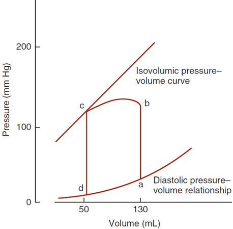
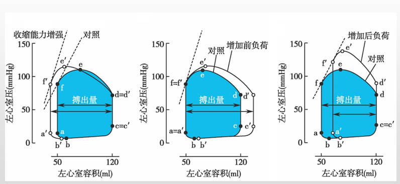
- normal pressure-volume loop of the left ventricle
- Cardiac function index
- 每搏输出量stroke volume
- the volume of blood ejected by a ventricle during each contraction.
- 𝑉𝑠 = 𝑉𝑒𝑑 −𝑉𝑒𝑠 = 125−55 =70𝑚𝑙
- Stroke Volume = End-Diastolic Volume - End-Systolic Volume
- 射血分数ejection fraction
- the fraction of the end diastolic volume that is ejected.
- 𝑉s/𝑉𝑒𝑑 = 55~65%
- 每分输出量cardiac output
- the volume of blood ejected by a ventricle each minute. 𝑉s×𝐻𝑅 = 4.5~6.0𝐿/𝑚𝑖𝑛
- 每搏输出量×心率
- 心指数cardiac index
- the cardiac output per square meter of body surface.
- 每搏功stroke work
- the amount of energy the ventricle converts to pump blood during each contraction.
- The work performed by a ventricle converts into
- ① potential energy that increases the blood pressure
- ② kinetic energy of the blood flow (only 1%)
- 𝑊 =𝑉𝑆×∆𝑃 + 1/2 ×𝑚𝑆×𝑣^2
- 𝑊𝐿𝑉 = 6×𝑊𝑅𝑉 左心室每博功是右心室的6倍
- 每搏输出量stroke volume
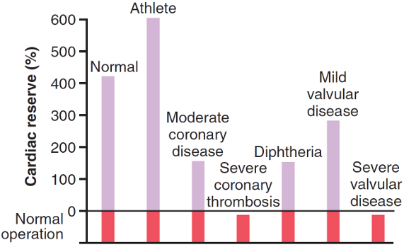
- Cardiac reserve
- At rest, cardiac output = 5
6L,maximum cardiac output =2530L - Cardiac reserve (心力储备) is the difference between a person’s maximum cardiac output and cardiac output at rest.
- includes
- stroke volume reserve (搏出量储备)
- Ved=125→140ml, Ves=55→20ml
- heart rate reserve (心率储备)
- 75→180 beats/min.
- However, heart rate over 180 beats/min shortens the diastole too much, leading to insufficient ventricular filling and decreased cardiac output
- stroke volume reserve (搏出量储备)
- 普通人的心力储备主要来源于心跳增加
- At rest, cardiac output = 5
- Factors controlling cardiac output
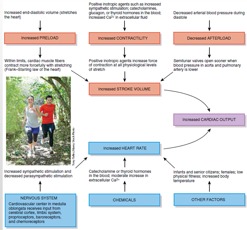
- 𝐶𝑎𝑟𝑑𝑖𝑎𝑐 𝑜𝑢𝑡𝑝𝑢𝑡 = V𝑠 × HR
- Factors controlling V𝑠
Vs = Ved - Ves- preload
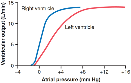
影响Ved- Preload = initial length = Ved → Ped = PA at atrial systole
刚开始收缩时候心室心房血压相等 - relaxed cardiac muscle is at less than optimum length
- Additional stretch therefore produces a significant increase in contraction force
- normal Ped = 5~6mmHg
- optimum Ped = 12~15mmHg
- When cardiac output is regulated by changes in cardiac muscle fiber length, it is referred to as heterometric regulation 异长自身调节
- Preload = initial length = Ved → Ped = PA at atrial systole
- afterload
影响Ves- Afterload is the resistance against which blood is expelled, namely the blood pressure in the aorta for the left ventricle
- In theory, an increase in afterload causes stroke volume to decrease
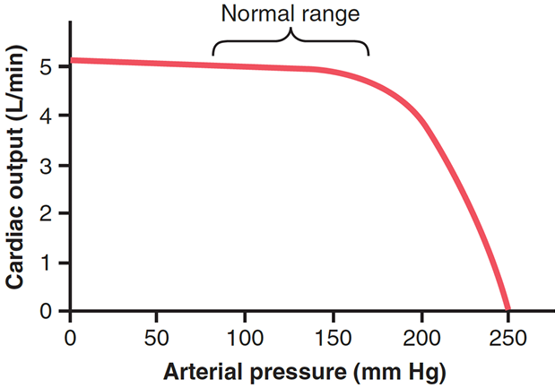
- In fact, increasing the arterial动脉的 pressure in the aorta does not decrease the cardiac output until the mean平均 arterial pressure rises above 160mmHg
- contractility
- Contractility is mainly determined by the numbers of activated cross-bridge and the enzyme activity of ATPase in myosin head
- Given a constant preload, the stroke volume can be changed by neural/humoral regulation.
- Effect on the strength of cardiac contraction is called inotropic action 变力作用
- preload
- Factors controlling HR
- Effect on the heart rate is called chronotropic action 变时作用
- 图
- 图2
- Internal anatomy of the heart
- Anatomy of the blood vessels
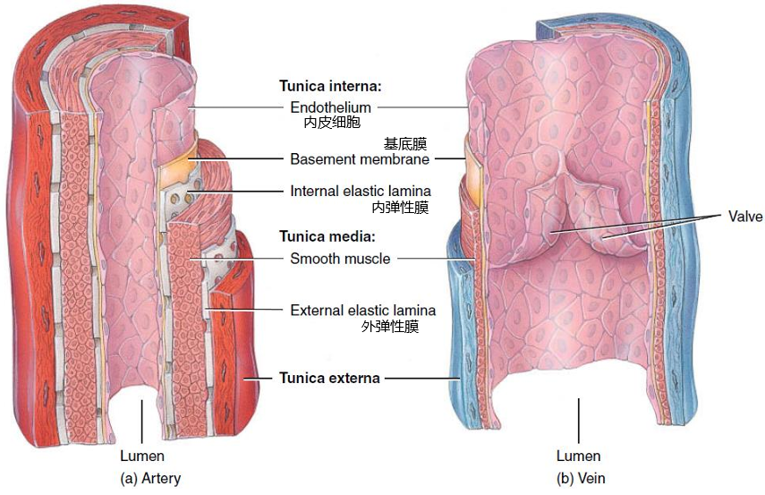
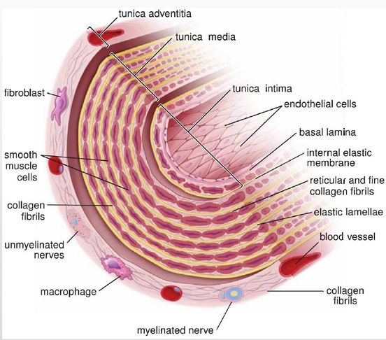
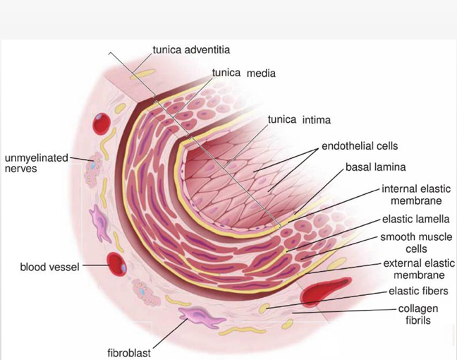
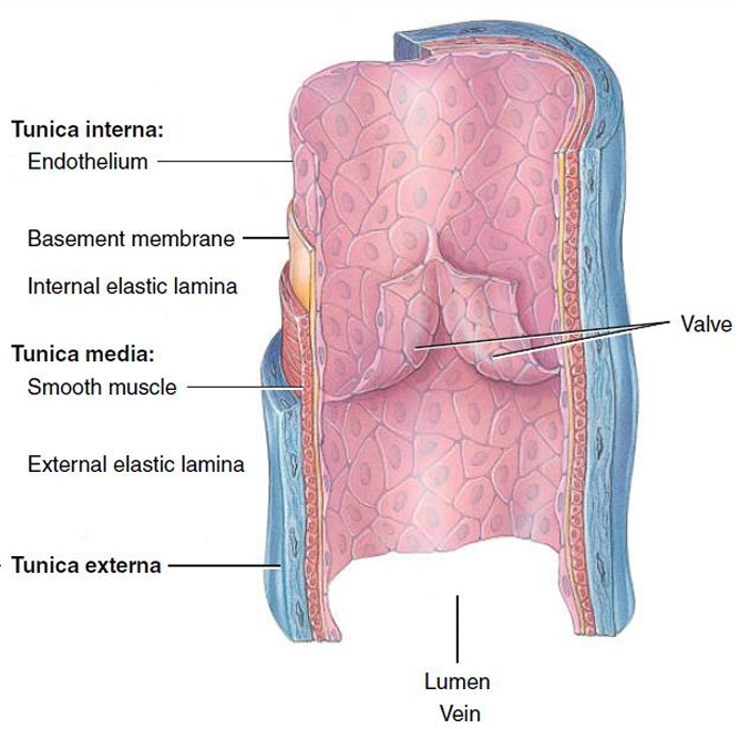
- 三层血管
- Tunica interna
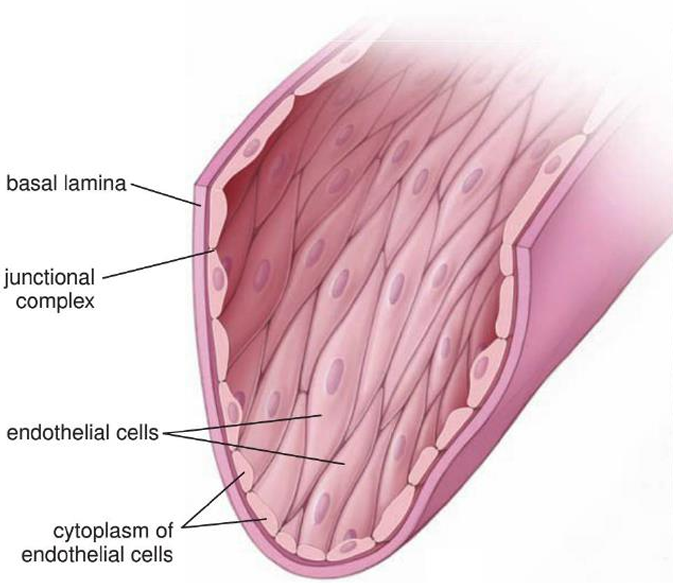
- Endothelium内皮细胞 is a layer of squamous epithelium.
- It forms the tunica interna with a basement membrane, and is in direct contact with the blood
- 图
- Tunica media
- The tunica media is a relatively thick layer comprising mainly smooth muscle and substantial大量的 amounts of elastic fibers
- Tunica externa
- The tunica externa is the outermost connective tissue layer, composed primarily of collagen fibers.
- It contains numerous autonomic nerves自主神经, tiny blood vessels called vasa vasorum (血管滋养管) that supply large vessels
- 图
- Tunica interna
- 不同类型的血管
- arteries
- Large arteries
elastic arteries, conducting arteries- have multiple sheets of elastic lamellae → elastic arteries
- elastic lamellae separates multiple layers of vascular smooth muscle
- Elastic arteries function as a pressure reservoir
- They conduct blood from the heart to medium arteries → conducting arteries
- Medium arteries
muscular arteries,distributing arteries- have more smooth muscle and less elastic lamellae → muscular arteries
- diatribute blood to different places → distributing arteries
- have more smooth muscle and less elastic lamellae → muscular arteries
- Small arteries and arterioles 小动脉
resistance arteries- Small arteries and arterioles are distinguished from one another by the number of smooth muscle cell layers in tunica media
- Because of their diameters, small arteries and arterioles produce a large resistance against the blood flow → resistance arteries
- Large arteries
- Capillaries
exchange vessels- 特点
- 80,000 kilometers of capillaries
- The endothelial cells form a tube just large enough to allow the passage of RBCs one at a time. In many capillaries, the lumen is so narrow that RBCs fold on themselves to pass through the vessel
- primary function is exchange of substances between the blood and the interstitial fluid → exchange vessels
- composed of only a single layer of endothelial cells and a basement membrane, lacking both a tunica media and a tunica externa
- 分类
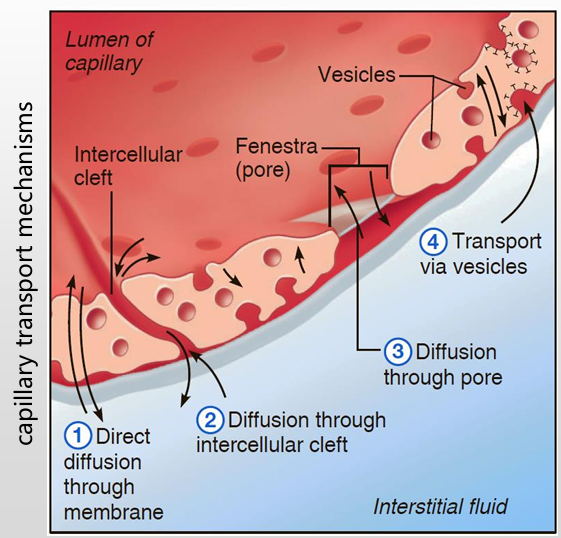
根据物质穿过的难易程度分类- Continuous capillaries
- The endothelial cells, held together by tight junctions, form a continuous tube.
- Cells are separated by narrow intercellular clefts (4nm), which hold back blood cells, platelets and large molecules like plasma protein.
- Continuous capillaries are found in the CNS中枢神经系统, the skin, lungs and muscles.
- Fenestrated capillaries
- The plasma membranes of endothelial cells in these capillaries have many fenestrations窗孔 (70-100nm)
- Fenestrated capillaries are found in sites of active secretion, absorption or filtration, such as the endocrine glands内分泌腺, intestinal tract肠道, and kidney
- Discontinuous capillaries
- Discontinuous capillaries (sinusoidal capillaries窦状毛细血管/sinusoids) have large openings in their endothelial cells that are separated by wide irregular intercellular gaps.
- Also, endothelial cells rest on a discontinuous basement membrane
- Proteins and even blood cells can pass through these capillaries
- These capillaries can be found in the liver肝脏, spleen脾脏 and red bone marrow红骨髓
- Continuous capillaries
- 特点
- Veins
capacitance vessels- relatively thin-walled and flaccid松弛, and expand easily to accommodate an increased volume of blood → capacitance vessels
- many contain venous valves that direct blood toward the heart
- arteries
- 三层血管
下半
- Vascular physiology血管生理
- Hemodynamics血流动力学
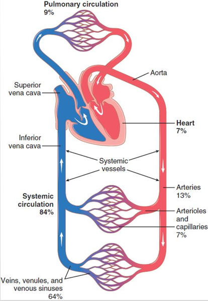
- 血液分布
- Blood flow 血流量
- the volume of blood that passes a given site in the circulation in a given time
- Poiseuille’s law
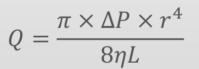
- Q流量
- r管道半径
- η粘度系数
- ΔP血管两侧压力差
- L管道长度
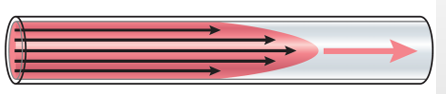
- laminar flow 层流
- blood flows at a steady rate through a long, smooth blood vessel, it flows in streamlines
- Poiseuille’s law can be applied to it
- blood flows at a steady rate through a long, smooth blood vessel, it flows in streamlines
- turbulent flow 湍流
- blood flows in all directions and continually mixes within the vessel
- Usually if Re>2000, turbulence will occur (v is velocity of blood flow, d is the vessel diameter, p is density, η is the viscosity)
- blood flows in all directions and continually mixes within the vessel
- Blood resistance
- Blood resistance is the opposition to blood flow due to friction between blood and the walls of blood vessels
- 𝑄 = ∆𝑃 / R
- 𝑅= 8𝜂𝐿 / π𝑟4
- R可以跟电路一样算串联并联
- 血液分布
- Arterial blood pressure动脉血压
- Blood pressure is the force exerted by blood against a vessel wall
- Systolic pressure 100
120mmHg, diastolic pressure 6080mmHg - 动脉血压的形成
- Sufficient blood filling in the circulatory system
- Ejection of blood by the heart
- Peripheral resistance
- The elastic distention of the aorta and large arteries
- 测量血压
- direct measurements
- indirect measurements
- Korotkoff sound method
- 血压的影响因素
- Stroke volume每搏输出量↑, arterial blood↑, pressure on the vessel wall↑, systolic pressure↑
- Heart rate↑, period of diastole↓, blood leaving the aorta during diastole↓, blood remaining in the aorta↑, diastolic pressure↑
- Peripheral resistance外周阻力↑, blood velocity during diastole↓,blood remaining in the aorta↑, diastolic pressure↑
- Arterial elasticity↓, blood pressure buffering↓, systolic pressure↑, diastolic pressure↓
- Blood loss↓, blood pressure↓
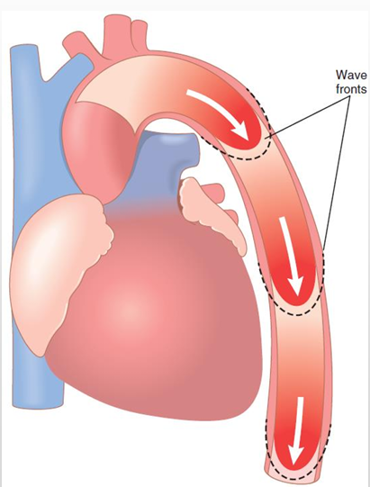
- Pressure pulse 脉搏
- Pressure pulse is a periodic change of the pressure and volume of the blood vessel
- 图像
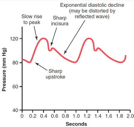
- The ascending limb - rapid ejection
- The descending limb - reduced ejection & diastole
- The dicrotic notch (降中峡) - blood backflows at the moment of closure of SL valves
- Transmission of the pressure pulse is a bit faster than the blood flow
- The greater compliance血管顺应性 of the each vascular segment, the slower the velocity
顺应性是指血管在压力作用下扩张的能力- 3~5m/s in the aorta
- 7~10m/s in large arteries
- 15~35m/s in small arteries
- Venous circulation
- The pressure in the venules is 12~18mmHg, and 0mmHg in the right atrium (veins have little resistance)
- Venous return is affected by
- ①mean filling pressure (平均充盈压)
- ②contractility of cardiac muscle
- ③ gravity
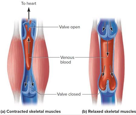
- ④skeletal muscle pump
- ⑤thoracic pump 胸腔泵
- Microcirculation
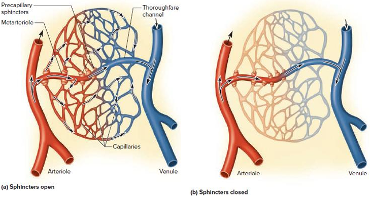
- The arterioles小动脉, capillaries and venules小静脉 form the miscrocirculation
- blood flows intermittently through capillaries due to vasomotion (血管运动), which may occur 5 to 10 times per minute
- 血管运动是括约肌间歇性收缩舒张,控制血流
- Capillary exchange
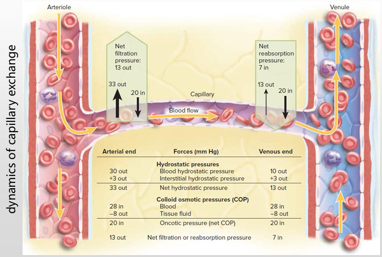
- 四种方法
- 判断水往哪流 +BP-HP-πB +血压-外面的水压-血液的渗透压(吸水)
- 四种方法
- Hemodynamics血流动力学
- Cardiovascular regulatory mechanisms
- Neural regulation
- afferent parts传入部分
- Baroreceptors 压力感受器
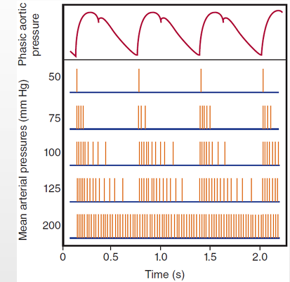
- detect blood pressure,High BP increases discharge rate of baroreceptors.
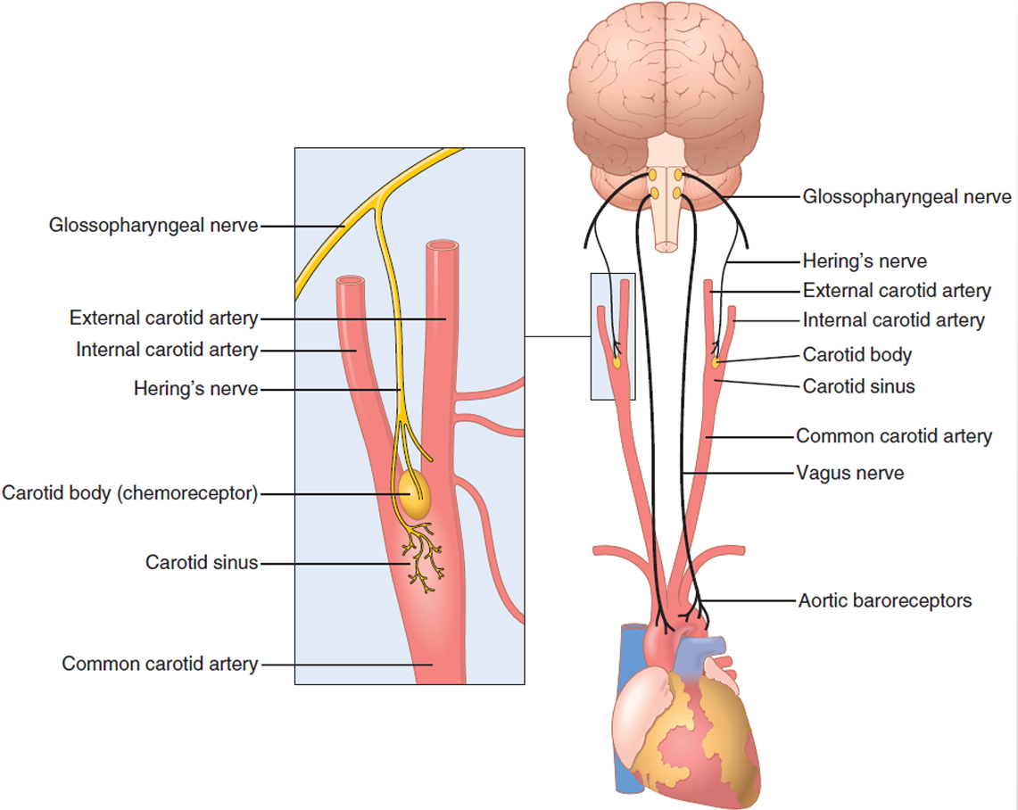
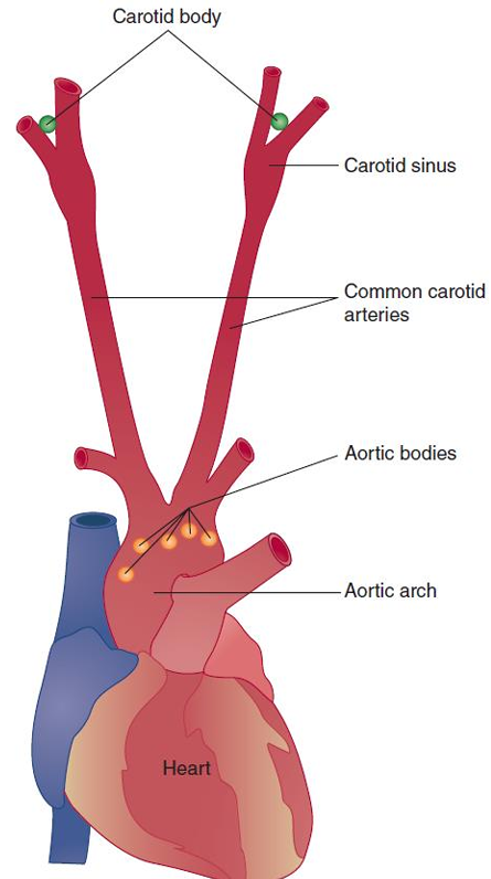
- 上面的是颈动脉窦,下面的是主动脉弓
- carotid sinus 颈动脉窦
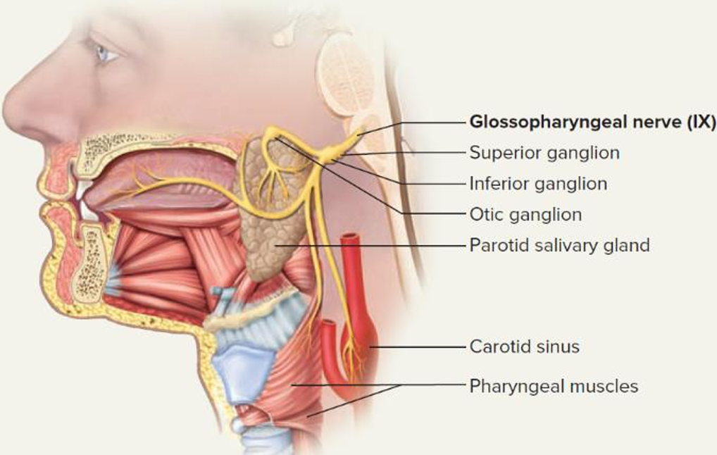
- carotid sinus 颈动脉窦 → carotid sinus nerve (Hering’s nerve) → glossopharyngeal nerve 舌咽神经 → cardiovascular center in the medulla oblongata 延髓
- carotid sinus 颈动脉窦 → carotid sinus nerve (Hering’s nerve) → glossopharyngeal nerve 舌咽神经 → cardiovascular center in the medulla oblongata 延髓
- aortic arch 主动脉弓
- aortic arch 主动脉弓 → depressor nerve (aortic nerve) → vagus nerve → cardiovascular center in the medulla oblongata 延髓
- detect blood pressure,High BP increases discharge rate of baroreceptors.
- Chemoreceptors 化学感受器
- detect O2, CO2 and H+in the blood
- 除了出发的位置不同,神经与压力感受器相同
- carotid bodies颈动脉体
- carotid bodies颈动脉体 → carotid sinus nerve (Hering’s nerve) → glossopharyngeal nerve → cardiovascular center in the medulla oblongata 延髓
- aortic bodies主动脉体
- aortic bodies主动脉体 → depressor nerve (aortic nerve) → vagus nerve → cardiovascular center in the medulla oblongata 延髓
- Cardiopulmonary receptors 心肺感受器
- in the walls of atria, ventricles and pulmonary veins detect stretches and chemicals
- Inputs from the receptors → vagus nerve → cardiovascular center
- Baroreceptors 压力感受器
- the center
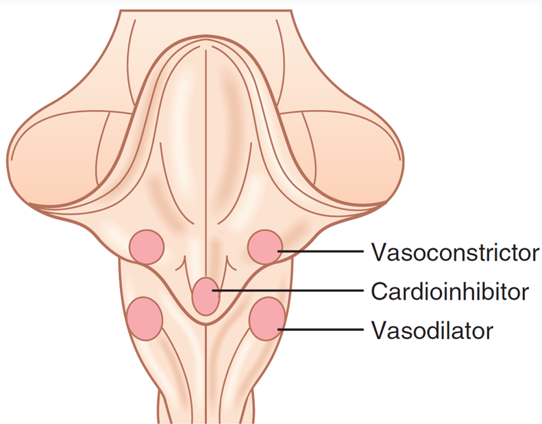
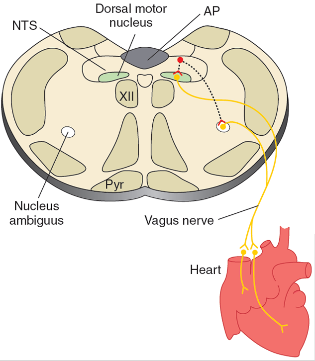
- The cardiovascular center is located in the medulla oblongata延髓 of brainstem脑干
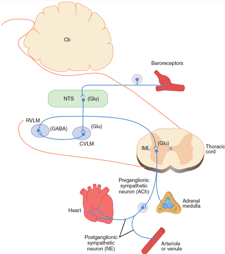
- The nucleus tractus solitaries (NTS, 孤束核) of medulla is a sensory area to receive signals from the vagus and glossopharyngeal nerves
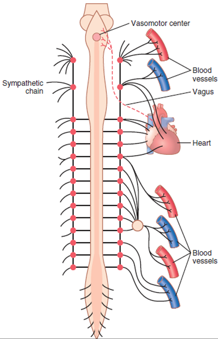
- vasoconstrictor(RVLM),vasodialator(CVLM),cardioinhibitor(nucleus ambiguus (疑核) and dorsal motor nucleus (背核)) 注意RVLM控制交感神经,可以控制心脏与血管,并不只是血管
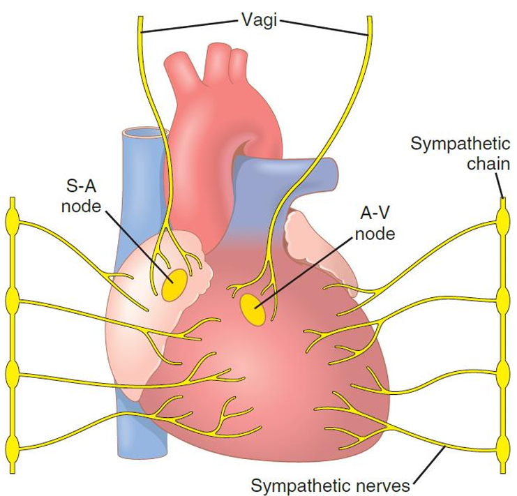
- 交感神经路径
- 迷走(副交感)神经路径
- 血压调控
- controlled by the sympathetic nerves only
- Receptors → NTS → CVLM -| RVLM→ sympathetic nerves
- 心脏调控
- controlled by the sympathetic and the vagus (parasympathetic) nerves
- Receptors → NTS → CVLM -| RVLM→ sympathetic nerves
- Receptors → NTS → nucleus ambiguus/dorsal motor nucleus → vagus nerves
- 交感神经控制大部分,迷走神经主要控制心房
- efferent parts
- sympathetic nerves
- The sympathetic nerves that innervate the heart start at T1~T5
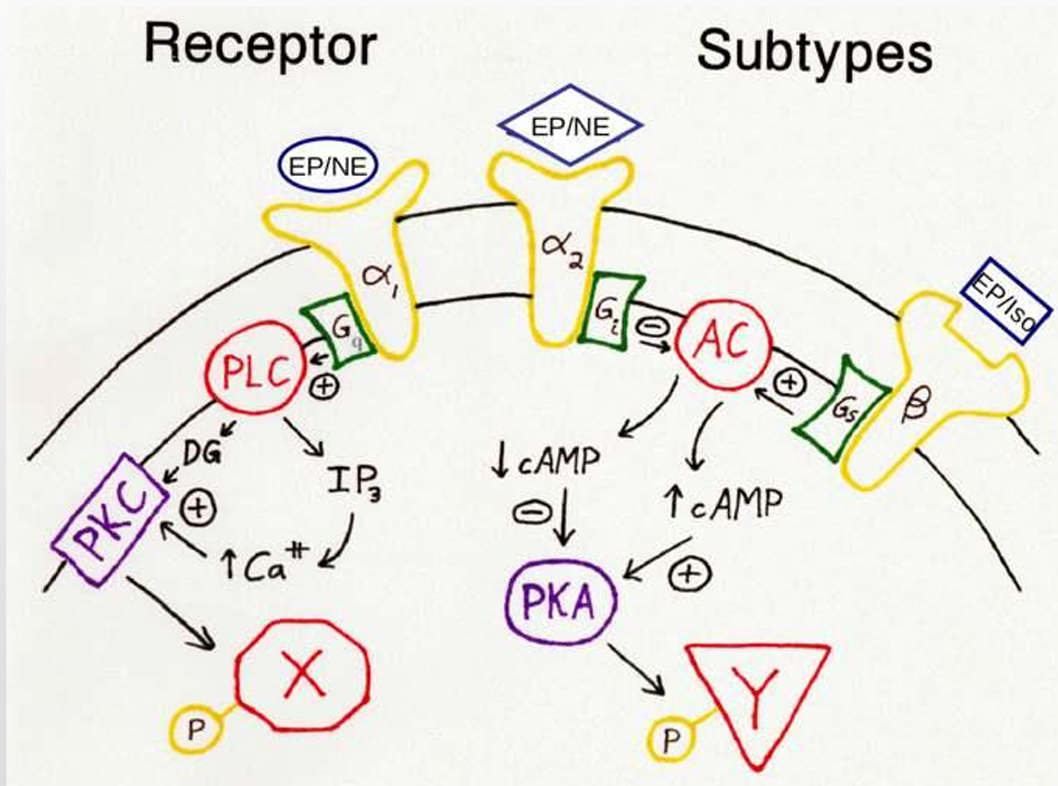
- When excited, these nerves release norepinephrine (NE, 去甲肾上腺素)
- NE 作用在β1-adrenoceptors of cardiac muscles与α1-adrenoceptors of vascular smooth muscles,看得出最后一个产生了PKA,一个产生了PKC
- Cardiac effects
- PKA phosphorylates ICa-L, ICa-T, If channels, and dissociate Ca2+ pump
- Working cells → ICa-L↑, Ca2+ pump↓, [Ca2+]i↑, contraction↑, positive inotropy (正性变力)
- Autorhythmic cells → If↑, ICa-T↑, ICa-L↑, spontaneous depolarization↑, heart rate↑, positive chronotropy (正性变时)
- Vascular effects
- [Ca2+]i↑, calmodulin activity↑, vasoconstriction↑, vessel diameter↓, vascular resistance↑, blood pressure↑
- The sympathetic nerves that innervate the heart start at T1~T5
- vagus nerves
- The vagus nerves start at the medulla
- When excited, the vagus nerves that innervate the atria release acetylcholine乙酰胆碱
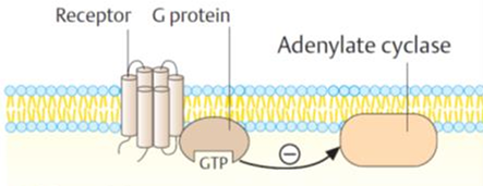
- Ach acts on the muscarinic (毒蕈碱型) receptors M2, inhibits the adenylate cyclase and reduce [cAMP]i
- Cardiac effects
- Phosphorylation of ICa-L, ICa-T, If channels weaken due to reduced PKA activity by [cAMP]i↓; IK-Ach is activated
- Working cells → ICa-L↓, [Ca2+]i↓, IK-Ach↑, repolarization↑, contraction↓, negative inotropy (负性变力)
- Autorhythmic cells → If↓, ICa-T↓, ICa-L↓, spontaneous depolarization↓, heart rate↓, negative chronotropy (负性变时)
- sympathetic nerves
- the reflex
- afferent parts传入部分
- Humoral regulation
- Epinephrine & norepinephrine 肾上腺素&去甲肾上腺素
- 被神经分泌就是神经递质,被肾上腺分泌就是激素
- adrenal medulla (肾上腺髓质)合成出来的80%是肾上腺素,20%是去甲肾上腺素
- 不同受体
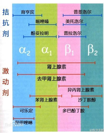
- α1
- in the vascular smooth muscle of skin, kidney and digestive tract
- vasoconstriction
- α2
- in the neurons
- β1
- in the cardiac muscle
- positive intropic and chronotropic effects
- β2
- in the skeletal muscle, and the smooth muscle of hepatic肝 vessels and respiratory tract 呼吸道
- vasodilation
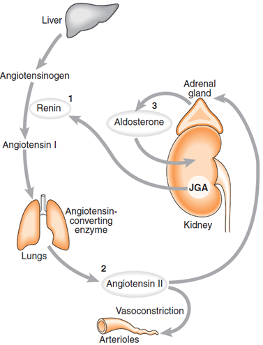
- Renin-angiotensin system 肾素-血管紧张素系统
- Vasoconstriction
- 机理
- 肾素是一种酶,可以水解angiotensinogen
- AngⅠ functions solely as the precursor of AngⅡ
- Angiotensin-converting enzyme (ACE) exists in the pulmonary vascular endothelium
- AngⅡ binds to AT1 receptors that activate Gq and Gi 最强大的收缩剂
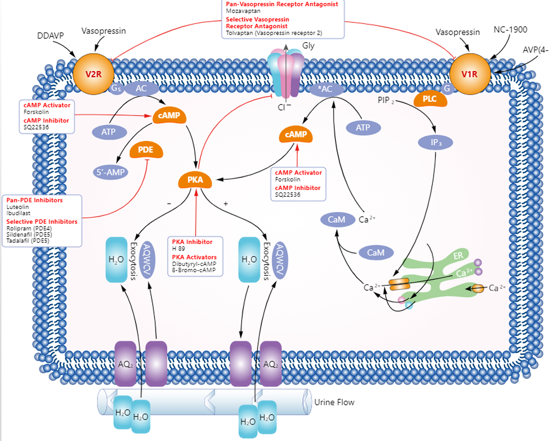
- Vasopressin 血管加压素
- VP is secreted by hypothalamus下丘脑
- It binds to V1 receptors of vascular smooth muscle
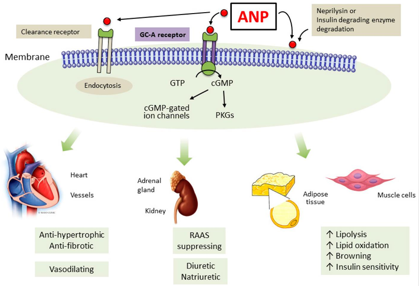
- Atrial natriuretic peptide 心房钠尿肽
- ANP is secreted by stretched atrial muscles
- ANP can strengthen Ca2+ pump and inhibit PLC
- vasodilation
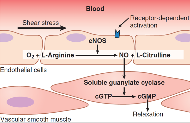
- NO
- 可以使cGMP↑,Ca2+↓
- vasodilation
- 可以使cGMP↑,Ca2+↓
- Epinephrine & norepinephrine 肾上腺素&去甲肾上腺素
- Neurohumoral regulation
- The neurohumoral regulation of cardiovascular system is a negative feedback control
- Changes of cardiovascular functions are eventually reflected by blood pressure
- Autoregulation
- Heterometric regulation
- 肌肉长度自动调节力度
- Metabolites
- for example, lactate and adenosine stimulate vasodilation
- Heterometric regulation
- Neural regulation
- Circulation through special regions
- Coronary circulation
- coronary冠状动脉连着主动脉刚出去的一部分
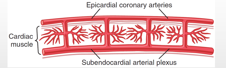
- diagram of the epicardial, intramuscular and subendocardial coronary vasculature 心外膜、心肌内和心内膜下冠状动脉血管分布的示意图
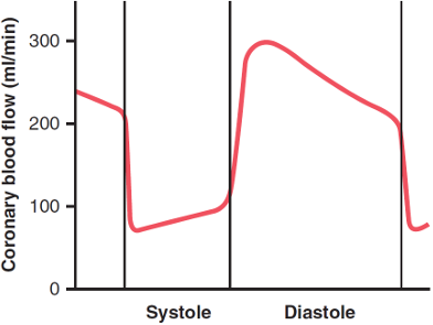
- phasic flow of blood through the coronary capillaries,血流量变化大,因为靠近心室
- coronary冠状动脉连着主动脉刚出去的一部分
- Pulmonary circulation 肺循环
- Low resistance
- low blood pressure
- low capacity
- Cerebral circulation 脑循环
- High blood flow and little change.
- Blood-brain barrier
- Coronary circulation


All articles in this blog are licensed under CC BY-NC-SA 4.0 unless stating additionally.