解剖学_腹膜&泌尿系统
腹膜&泌尿系统
- Peritoneum 腹膜
- 基础介绍
- Gr. Stretched round
- 定义 - Serous membrane浆膜 that stretches around/lines the abdominal cavity
- 作用 - Makes serous fluid
- 组织构成 - Made of mesothelium tissue 间皮//一种上皮组织
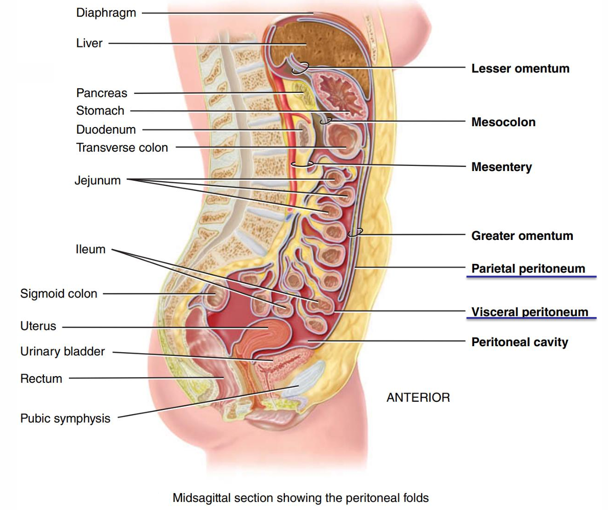
- parietal peritoneum 壁腹膜
- lines the wall of the abdominal cavity
- visceral peritoneum 脏腹膜
- covers the organs in the abdominal cavity
- 图
- Space in between is peritoneal cavity腹膜腔, which contains lubricating fluid.
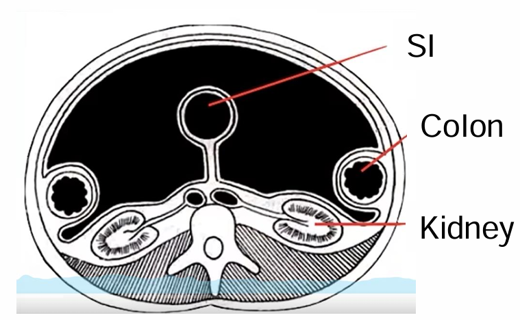
- The GI tract pushes in the peritoneum. 消化道是向内推入腹膜中的
- Peritoneum contains large folds大型皱襞, that bind the organs to one another and to the walls of the abdominal cavity.
- Major peritoneal folds
- The greater omentum 大网膜
- The lesser omentum 小网膜
- The mesentery 肠系膜
- The mesocolon 结肠系膜
- peritoneal cavity 腹膜腔
- space between the parietal and visceral peritoneum
- contains serous fluid
- 病-Ascites腹水肿
- accumulation of several liters of fluid
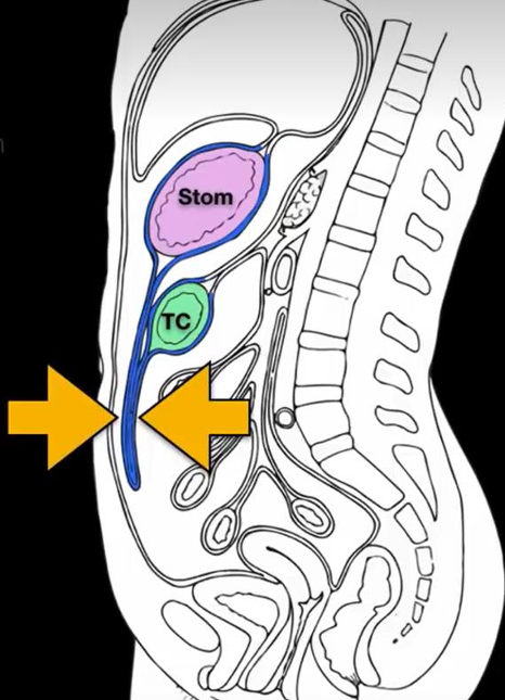
- greater omentum 大网膜
- attaches the stomach and transverse colon
- 4 layers
- 图
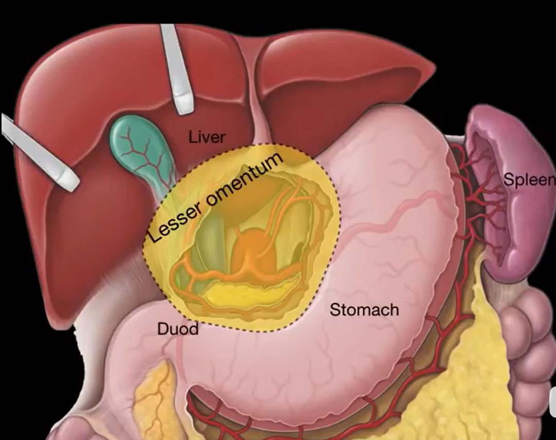
- lesser omentum 小网膜
- attaches to the stomach, duodenum and liver
- lesser omentum = Hepatogastric ligament + Hepatoduodenal ligament
- 图
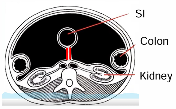
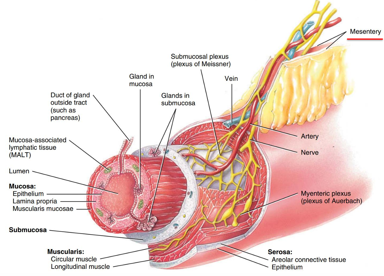
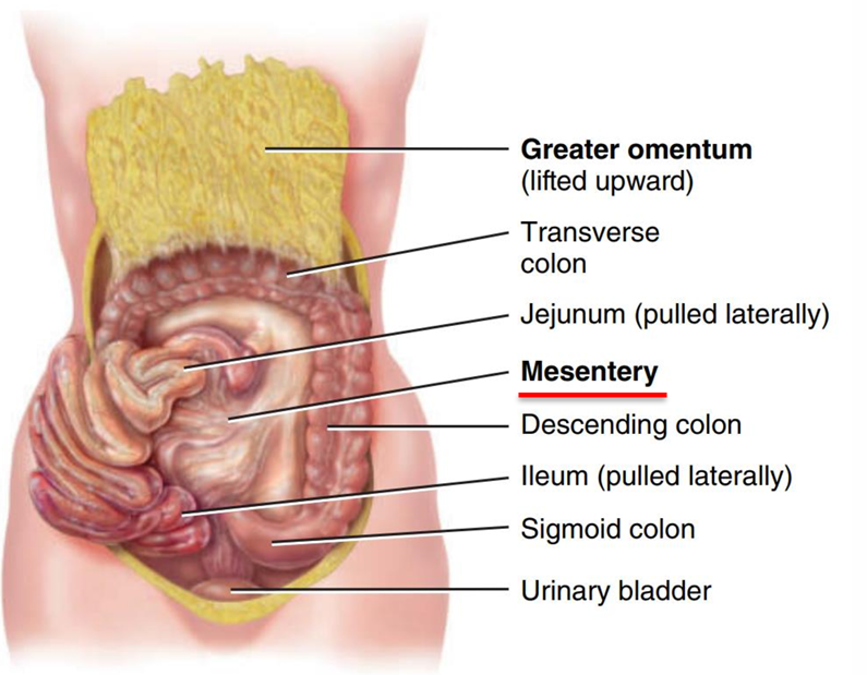
- mesentery 肠系膜
- portion of peritoneum//红色部分
- attaches small intestine (jejunum and ileum) to posterior abdominal wall
- double layers
- Blood vessels and nerves go through the double layers of the mesentery to supply the abdominal organs
- 连在小肠后面//大网膜提溜起来,小肠扒拉开
- portion of peritoneum//红色部分
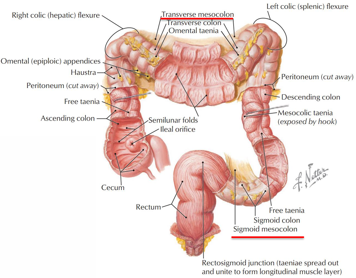
- mesocolon 结肠系膜
- Two separate peritoneum
- transverse mesocolon (transverse colon )
- sigmoid mesocolon (sigmoid colon)
- bind the transverse colon and sigmoid colon to the posterior abdominal wall
- Two separate peritoneum
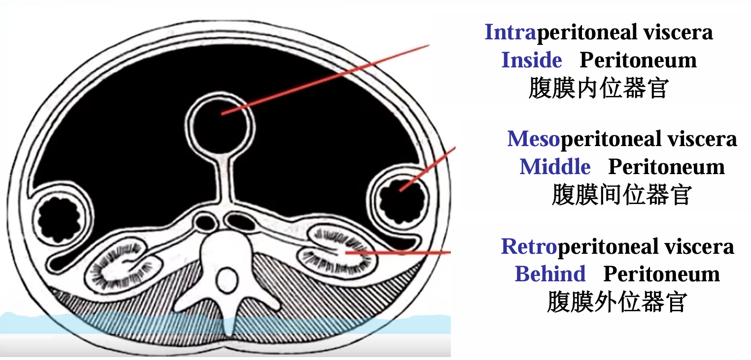
- 器官根据与腹膜相对位置的分类
- Intraperitoneal viscera 腹膜内位器官
- Stomach
- Duodenum (1st part)
- Jejunum and ileum
- Cecum
- Appendix
- Transverse colon
- Sigmoid colon
- Upper rectum
- Liver Pancreas (only the tail)
- Spleen
- Mesoperitoneal viscera 腹膜间位器官
- Ascending and descending colon
- Middle rectum
- Gallbladder
- Retroperitoneal viscera 腹膜外位器官
- Esophagus
- Duodenum (2nd-4th parts)
- Pancreas (head, neck and body)
- Kidneys
- Ureters
- Urinary bladder
- 图
- Intraperitoneal viscera 腹膜内位器官
- 基础介绍
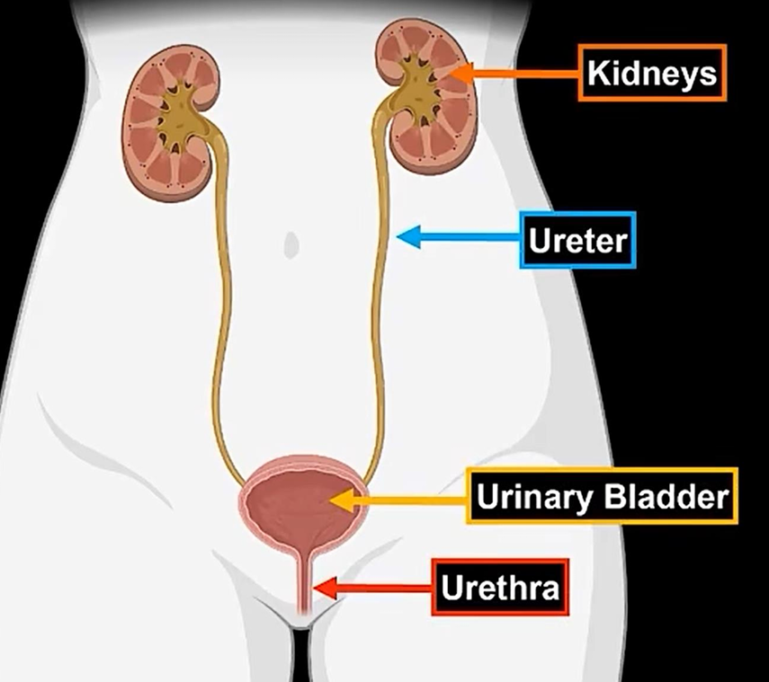
- Urinary System
- 构成
- Kidneys 肾
- Ureters 输尿管
- Urinary bladder 膀胱
- Urethra 尿道
- 图
- kidney
- Location
- Right kidney is slightly lower than the left because the liver occupies considerable space on the right side superior to the kidney
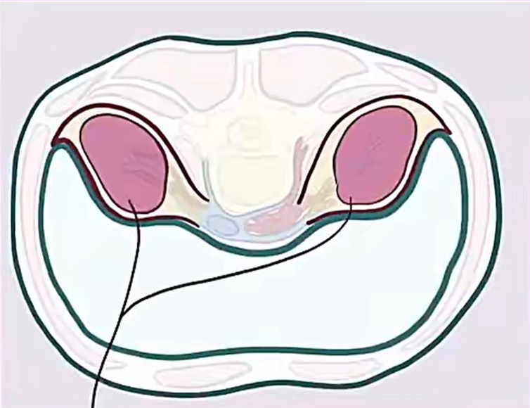
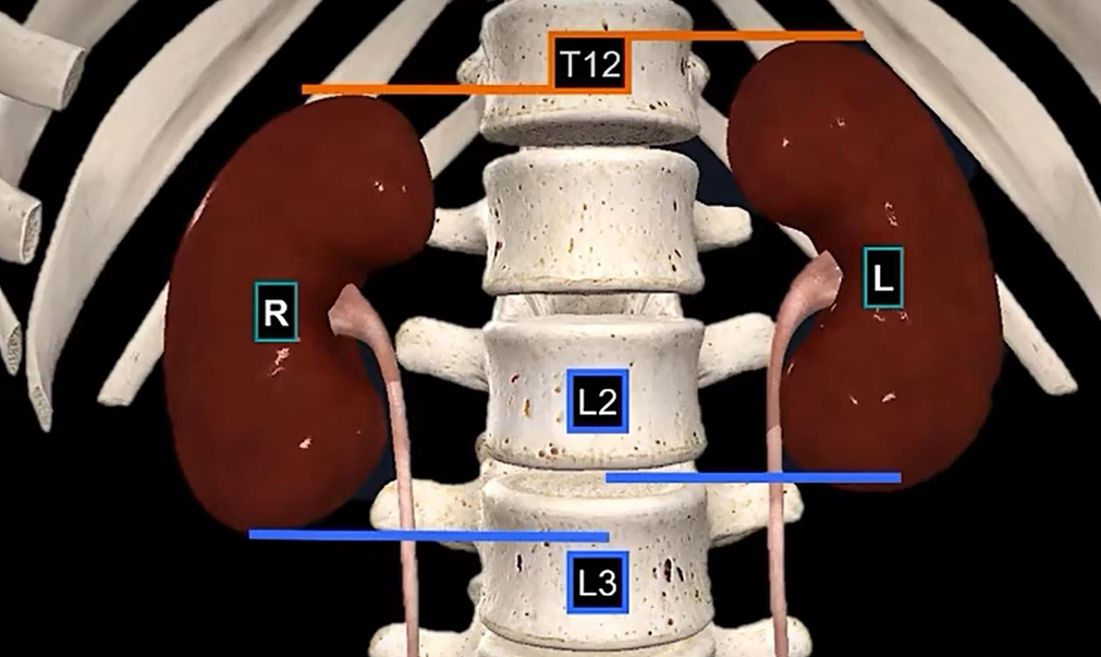
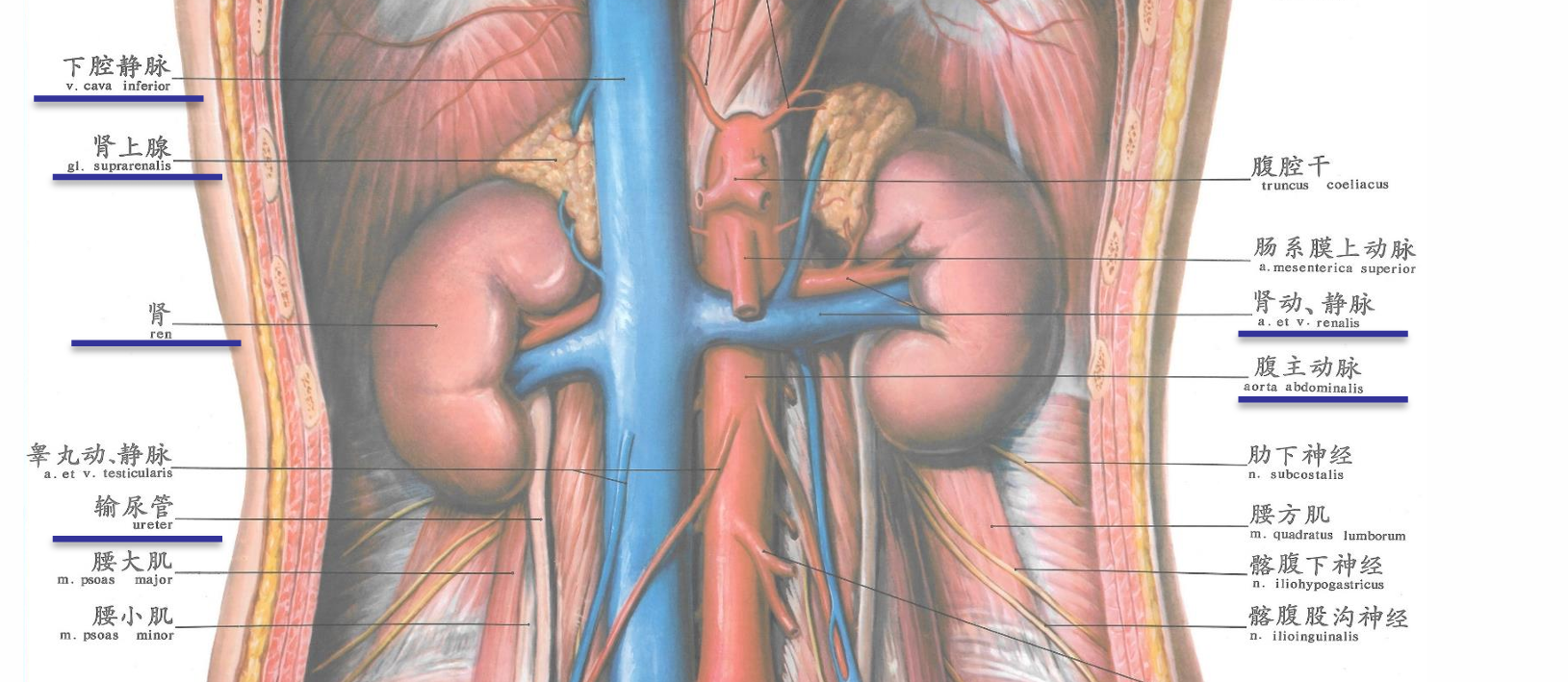
- Kidneys are retroperitoneal, along side the vertebral column
- locate between the levels of T12 and L2/L3
- partially protected by ribs 11 and 12
- External Anatomy of the Kidneys
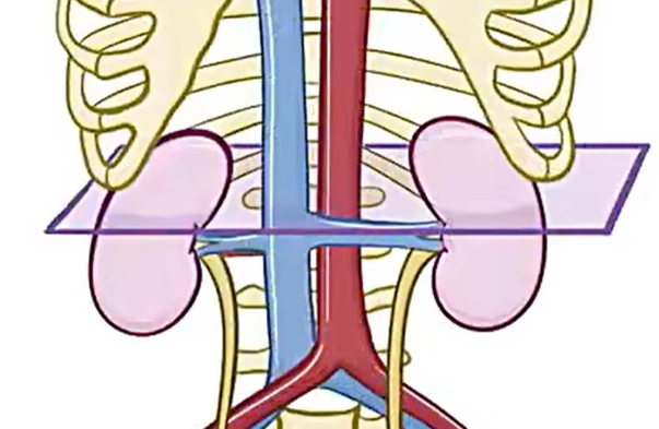
- Near the center of the concave border is the renal hilum肾蒂
- Entry & Exit for
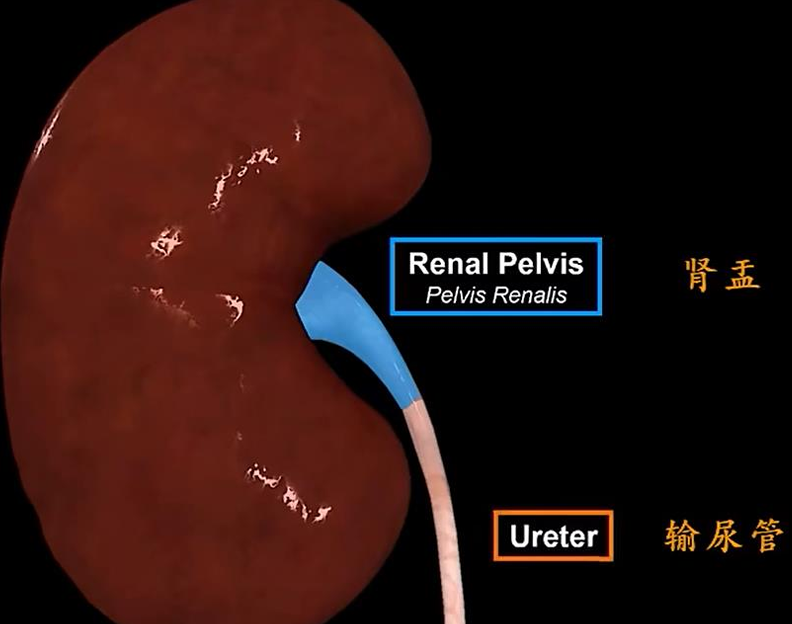
- Renal pelvis 肾盂
- Renal A
- Renal V
- lymphatic vessels
- nerves
- Renal pelvis 肾盂
- Suprarenal gland 肾上腺
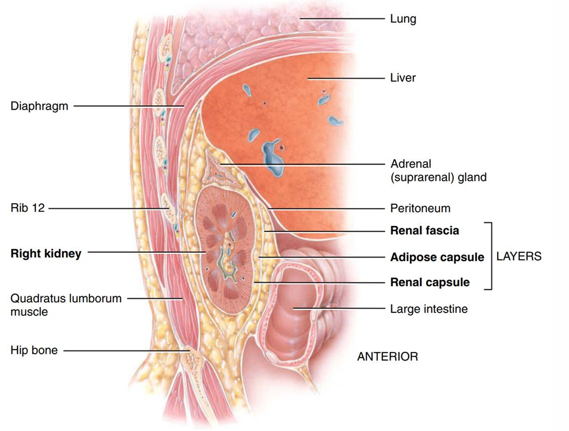
- Three layers of tissue surround each kidney
- deep layer - the renal capsule 纤维囊
- a smooth, transparent sheet of dense connective tissue
- gives kidney its distinctive shape
- middle layer - the adipose capsule 脂肪囊
- a mass of fatty tissue
- protects the kidney
- superficial layer - the renal fascia 肾筋膜
- a thin layer of dense connective tissue
- anchors the kidney to the surroundings
- 竖着劈开,最左边的是肋骨
- deep layer - the renal capsule 纤维囊
- Internal Anatomy of the Kidneys
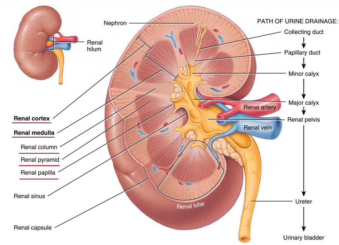
- a superficial, light red region called renal cortex 肾皮质
- a deep, darker reddish-brown inner region called renal medulla 肾髓质
- renal columns 肾柱,皮质向髓质延伸的部分,填充在肾锥体之间的空隙中
- medulla made of 10-18 renal pyramids 肾锥体
- apex (tip) of the renal pyramid called renal papilla 肾乳头
- 图
- 尿液汇集
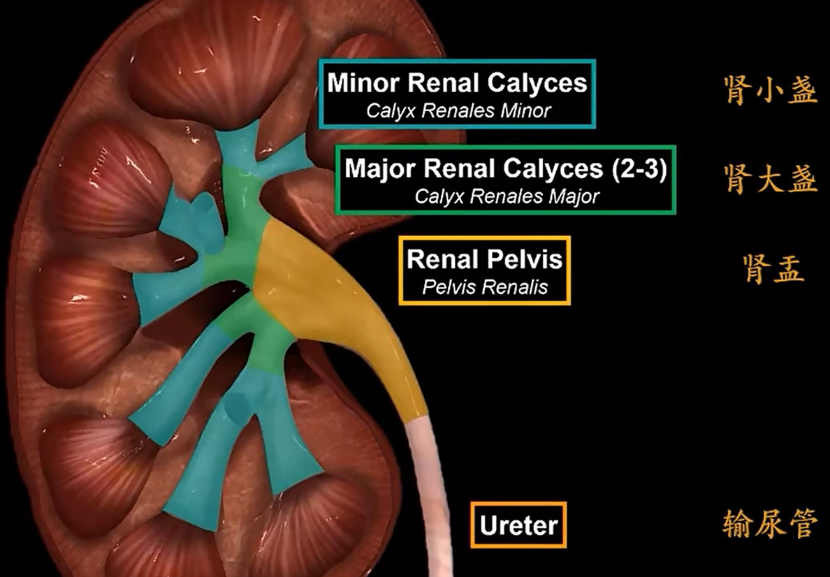
- 希腊语calyces = cups, singular calyx
- 1-3 papillary ducts → 1 minor calyx
- 2-3 minor calyces → 1 major calyx
- renal pelvis肾盂 is a single large cavity
- The hilum expands into a cavity within the kidney called the renal sinus
- Adipose tissue脂肪组织 helps stabilize the position of these structures in the renal sinus.
- 图
- Functions of the Kidneys
- Filter blood Kidneys filter 150 L of blood a day
- If 5L blood in body that means Entire volume of blood is filtered 30 times a day
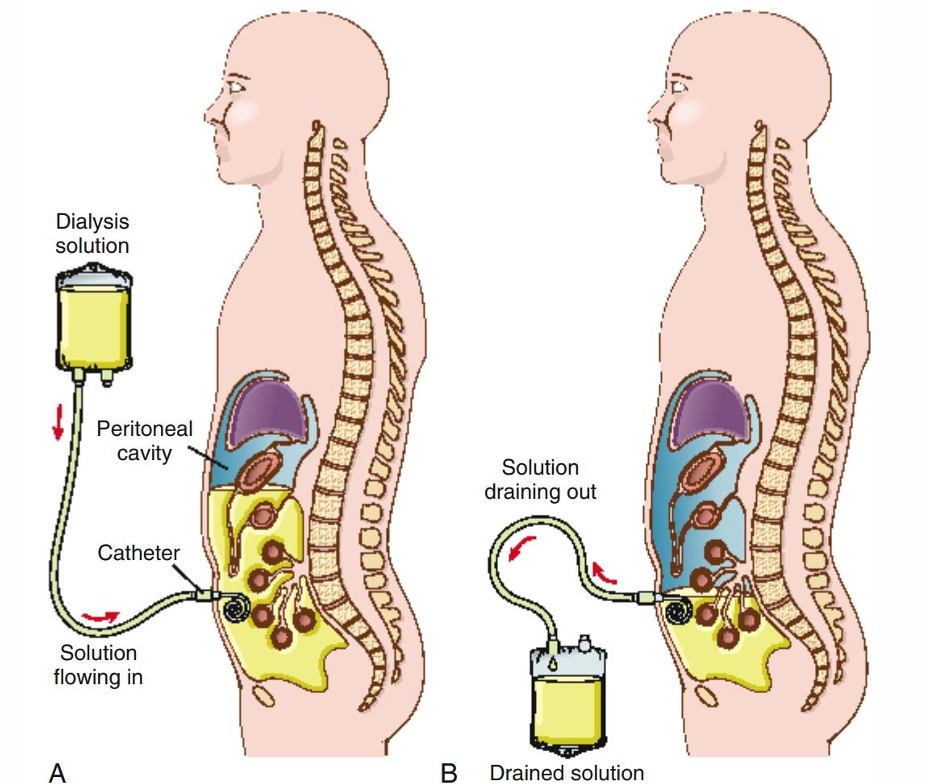
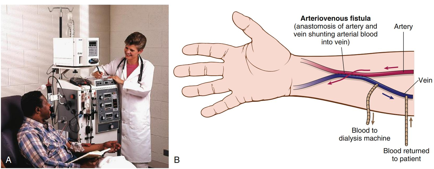
- 病-Peritoneal dialysis for chronic kidney disease慢性肾脏病的腹膜透析
- uses a catheter to introduce fluid into the peritoneal cavity
- wastes in the capillaries of the peritoneum pass out of the bloodstream and into the fluid
- fluid (with wastes) removed by catheter
- 将透析液注入腹腔,利用腹膜毛细血管与透析液之间的渗透/扩散作用净化血液
- 图
- 病-Hemodialysis透析
- filters blood through an artificial porous membrane
- returns the dialyzed blood to the patient’s body
- 图
- Location
- Ureter
- about 25 cm long
- retroperitoneal position
- 3部分
- Abdominal part 腹部
- extends from the renal pelvis to the iliac A 髂qia动脉
- Pelvic part 盆部
- After crossing the pelvic brim骨盆缘, the ureter enters the pelvic cavity towards the bladder.
- Intramural part 输尿管壁内部
- short segment that passes through the wall of the urinary bladder
- 1.5 cm
- Abdominal part 腹部
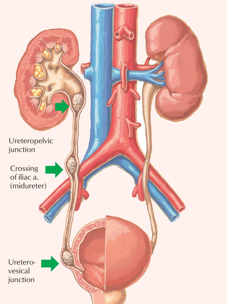
- The ureter has 3 constrictions
- Junction where the renal pelvis connects the ureter
- Point where the ureter crosses the iliac A (either the right or left) 髂动脉
- Junction where the ureter enters the urinary bladder
- 这三处位置容易出现renal stone
- Urinary Bladder
- In the pelvic cavity
- a muscular “bag” servers as a reservoir for the urine
- holds up to 800 mL of urine
- 位置
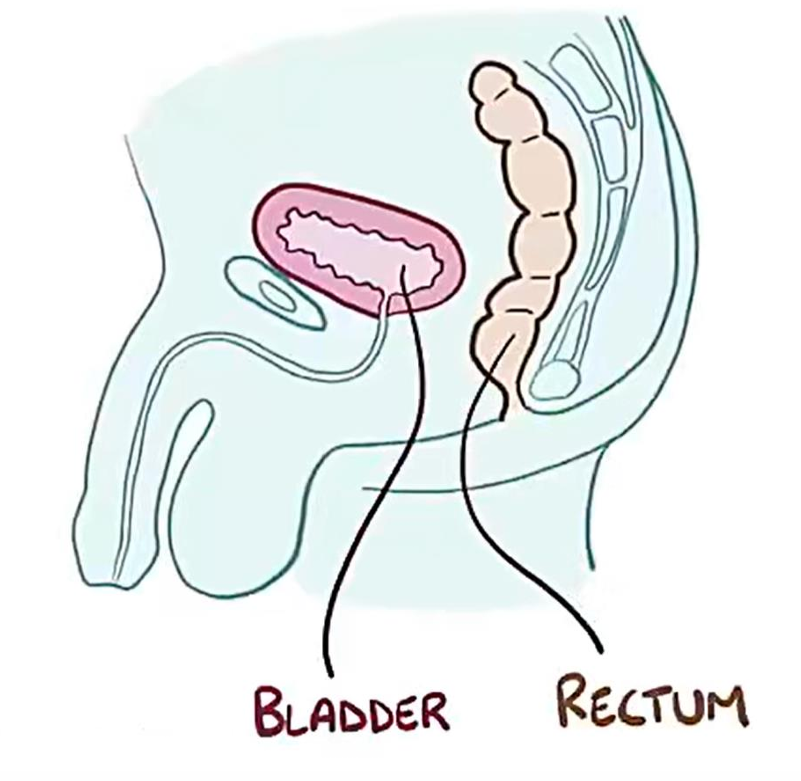
- In males, it is directly anterior to the rectum
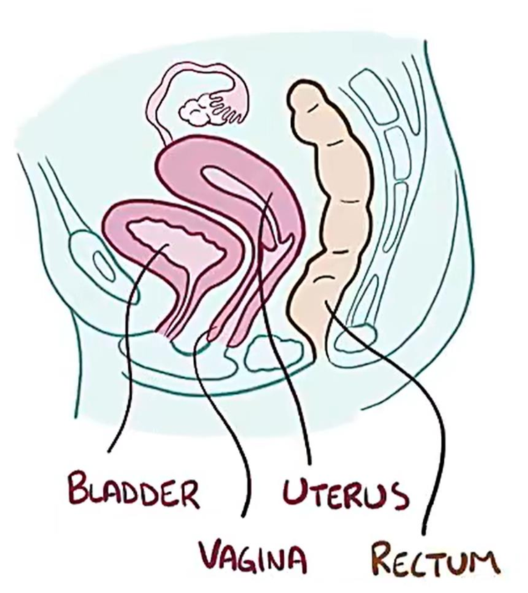
- In females, it is anterior to the vagina and inferior to the uterus
- In males, it is directly anterior to the rectum
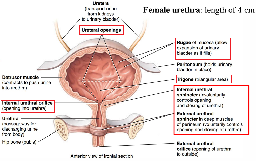
- Internal anatomy of the urinary bladder
- Trigone
- In the floor of the urinary bladder
- Two corners - ureteral openings
- Third corner - opening into urethra 尿道内口
- Mucosa is firmly bound to the muscularis - smooth appearance
- Sensitive to expansion
- Muscularis consists of three layers of smooth muscle
- inner longitudinal fibers
- middle circular fibers - form an internal urethral sphincter 尿道内括约肌
- outer longitudinal fibers
- external urethral sphincter尿道外括约肌 - composed of skeletal muscle
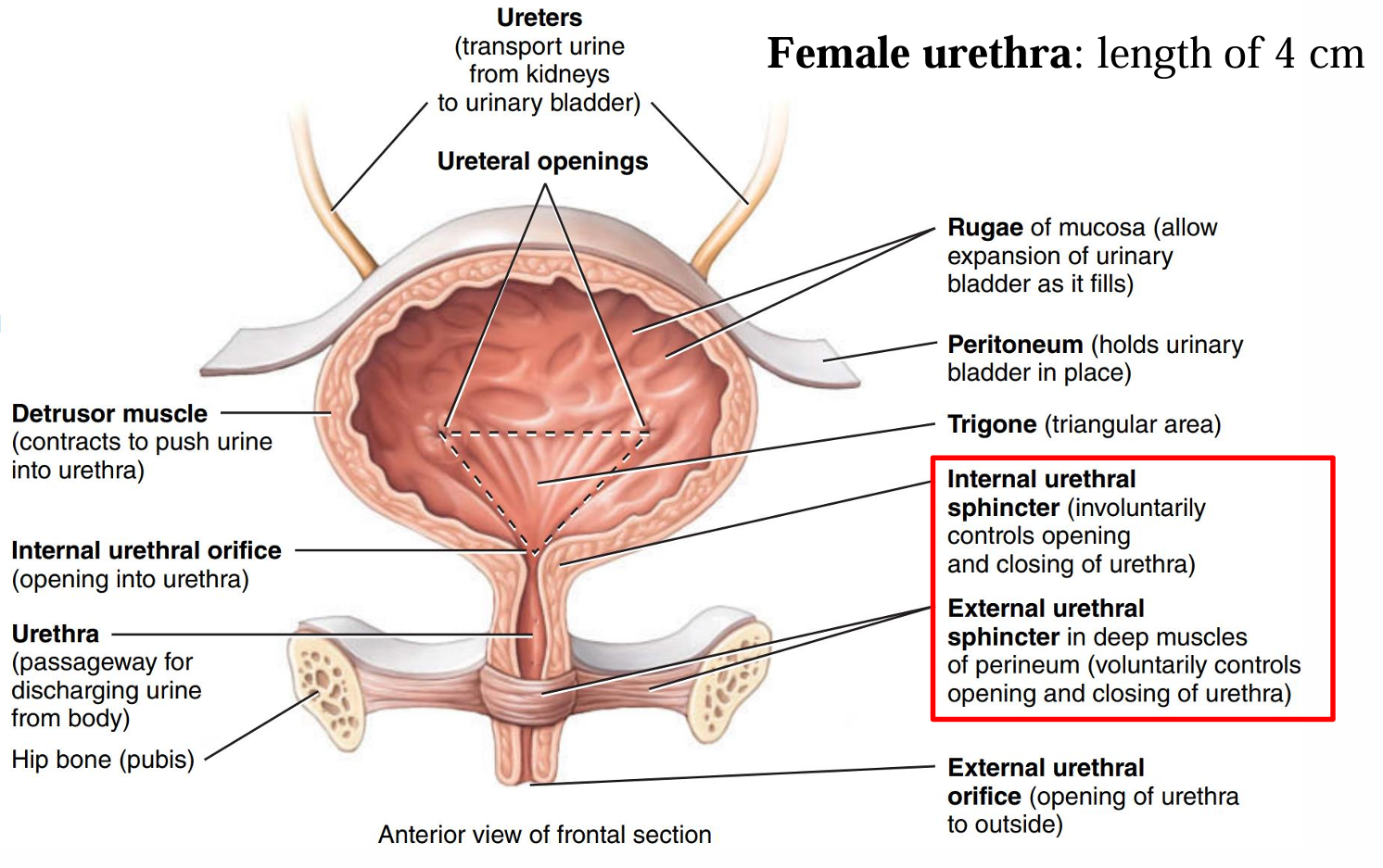
- 图//女性
- Trigone
- Urethra
- 女性
- length of 4cm
- length of 4cm
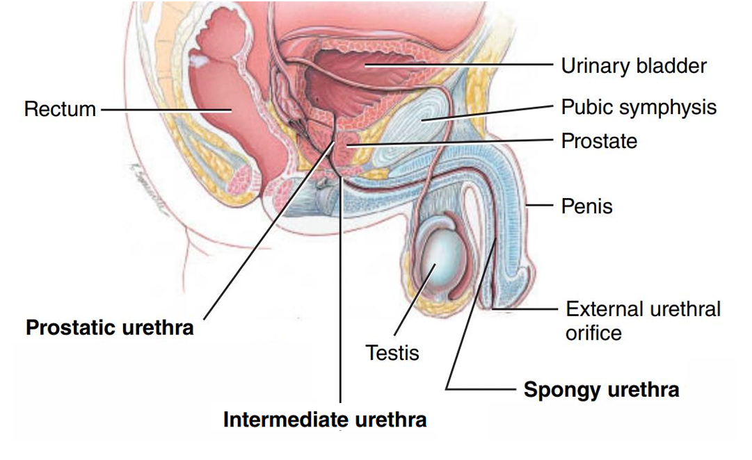
- 男性
- length of 20 cm
- 3部分
- prostatic urethra 前列腺部
- passes through the prostate
- membranous urethra/intermediate urethra 膜部
- passes through the deep muscles of the perineum 会阴尿道外括约肌
- spongy urethra 海绵体部
- passes through the penis
- 图
- prostatic urethra 前列腺部
- Urethral sphincters
- Internal sphincter
- smooth-muscle involuntary sphincter at the neck of the bladder
- External sphincter
- skeletal muscle voluntary sphincter surrounding the membranous urethra
- Internal sphincter
- males vs. females
- The urethra is 5 times longer in males
- The urethra is divided into 3 segments in males, but is only 1 short tube in females
- The urethra is a common duct for the urinary and reproductive systems in males.
- These two systems are separate in female.
- 女性
- 构成
All articles in this blog are licensed under CC BY-NC-SA 4.0 unless stating additionally.