解剖学_心血管
心血管
- heart
- Location
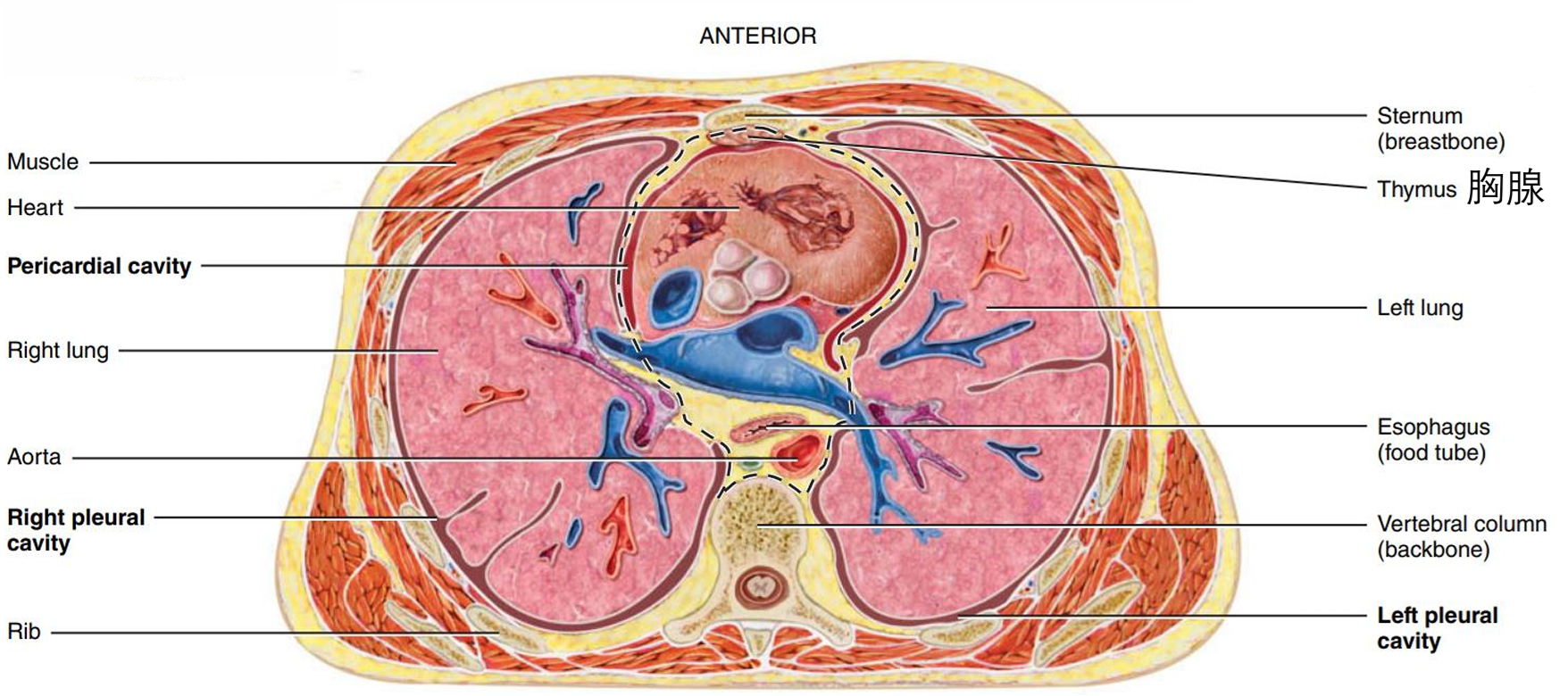
- In the mediastinum
- Between the lungs
- Mediastinum 纵隔 is a region that extends from sternum to vertebral column and from 1st rib to diaphragm
- 2/3 of the mass of the heart lies to the left of the body’s midline.
- Visualizes as a cone lying on its side
- In the mediastinum
- 外观
- Apex
- formed by the tip of LV and rests on the diaphragm
- directed anteriorly, inferiorly, and to the left
- Base
- opposite the apex
- formed by the atria, mostly the LA
- Anterior surface
- deep to the sternum and ribs
- Inferior/diaphragmatic surface
- rests on the diaphragm
- Right surface
- faces the right lung
- Left surface
- faces the left lung
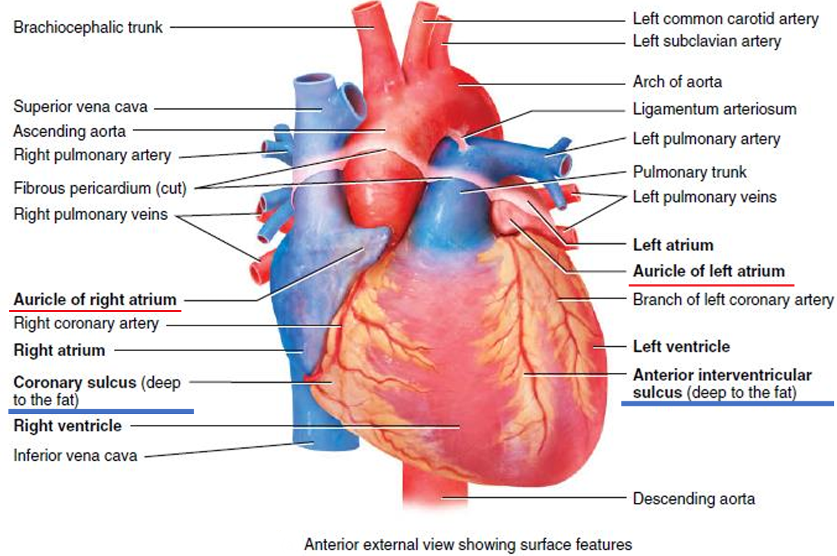
- Auricle 心耳
- resembles a dog’s ear
- on the anterior surface of each atrium
- Sulci
- a series of grooves沟 on heart surface
- contain coronary blood vessels and fat
- external boundary for 4 chambers
- coronary sulcus 冠状沟
- 分隔 atria and ventricles
- anterior interventricular sulcus 前室间沟
- 分隔 RV and LV on the anterior surface
- posterior interventricular sulcus 后室间沟
- 分隔 RV and LV on the posterior surface
- coronary sulcus 冠状沟
- 图
- Apex
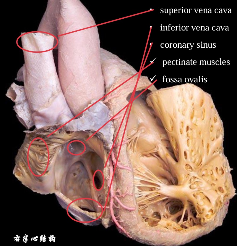
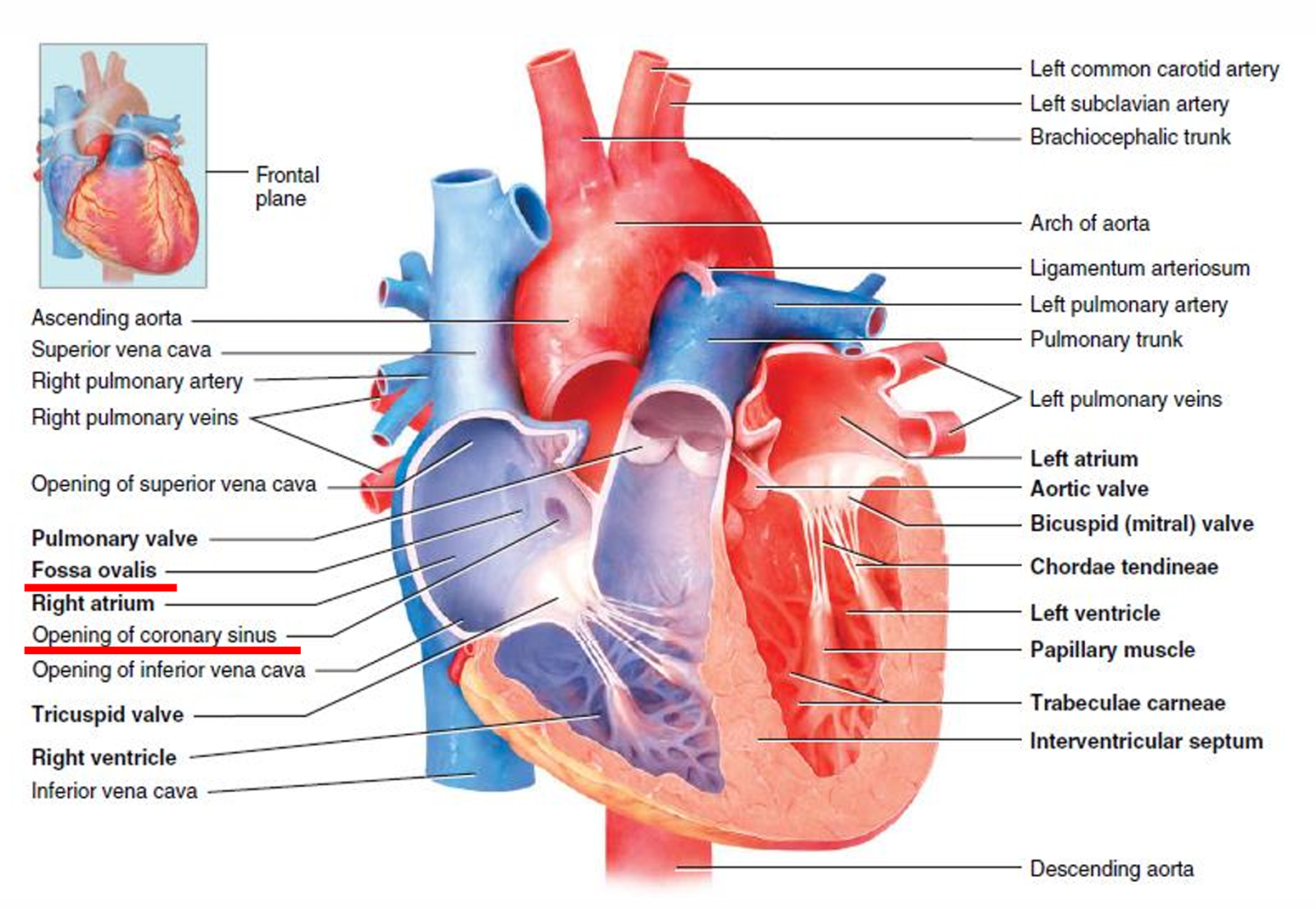
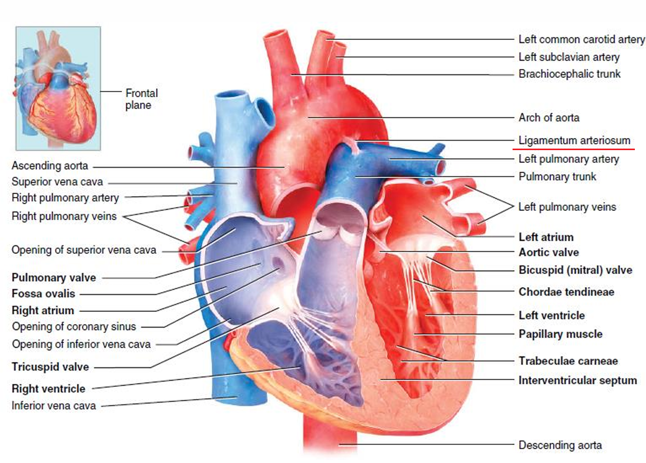
- Right atrium
- Entry
- Superior vena cava
- Inferior vena cava
- Coronary sinus 冠状窦
- opening of deoxygenated blood from the myocardium
- Exit
- Tricuspid valve
- 结构
- Smooth posterior wall of the inner layer
- Rough anterior wall of the inner layer due to pectinate muscles 梳状肌//左边掀起来的
- Pectinate muscles extend into the auricle
- Interatrial septum (septum = a dividing wall)
- fossa ovalis - oval depression 卵圆窝
- 图
- Entry
- Right ventricle
- Exit
- pulmonary valve into pulmonary trunk 肺动脉干
- pulmonary trunk divides into right and left pulmonary arteries
- 结构
- forms most of the anterior surface of the heart
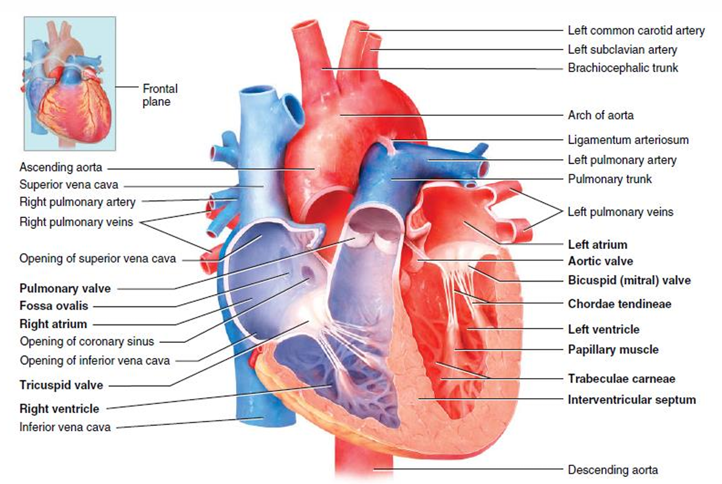
- inside of the RV contains trabeculae carneae 肉柱
- cusps of the tricuspid valve connect to chordae tendineae 腱索
- chordae tendineae connect to papillary muscles 乳头肌
- RV separated from LV by interventricular septum
- 图
- Exit
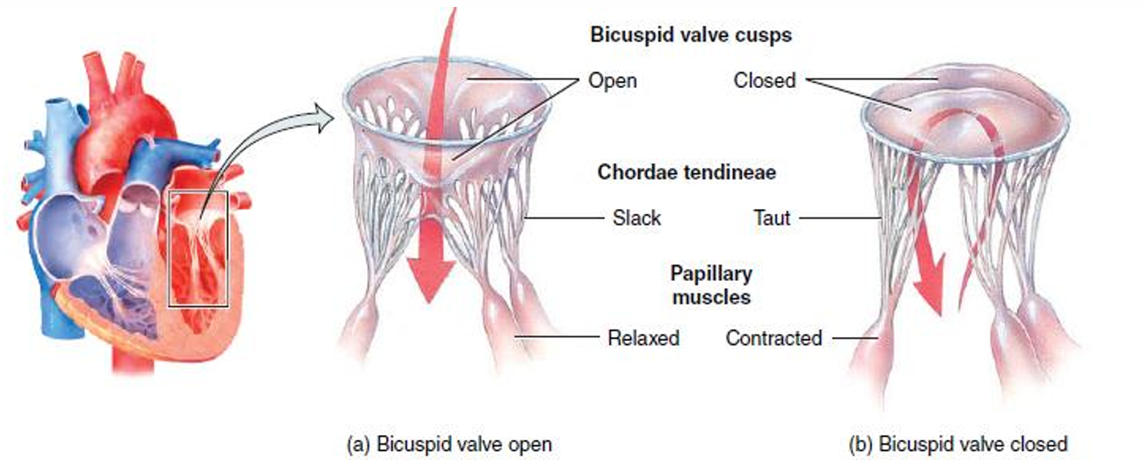
- Operation of tricuspid and bicuspid
- ventricles relax → papillary muscles relax → chordae tendineae are slack → blood flows from a higher pressure in the atria to a lower pressure in the ventricles through open valves
- ventricles contract → pressure of the blood drives the cusps upward and close the opening → papillary muscles contract → pulls on chordae tendineae → prevents the backflow of blood
- 图
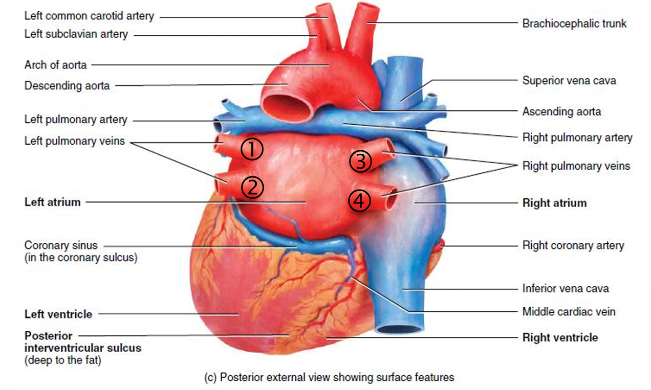
- Left atrium
- Entry
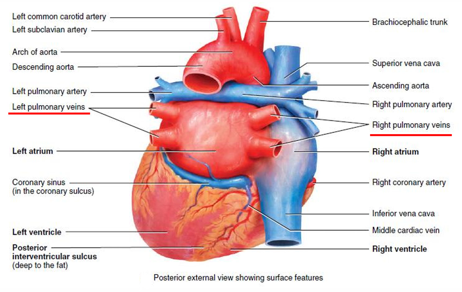
- four pulmonary veins
- Exit
- bicuspid valve
- forms most of the base of the heart
- smooth inner layer as pectinate muscles only in the auricle of the LA
- 后面看, 左右肺静脉流入左心房
- Entry
- Left ventricle
- Thickest chamber
- Forms the apex of the heart
- Contains trabeculae carneae and chordae tendineae
- Exit
- aortic valve into the ascending aorta
- ascending aorta
- arch of the aorta
- descending aorta (thoracic aorta and abdominal aorta)
- aortic valve into the ascending aorta
- ligamentum arteriosum 动脉韧带 connects the pulmonary trunk and the aorta
- Fibrous rings
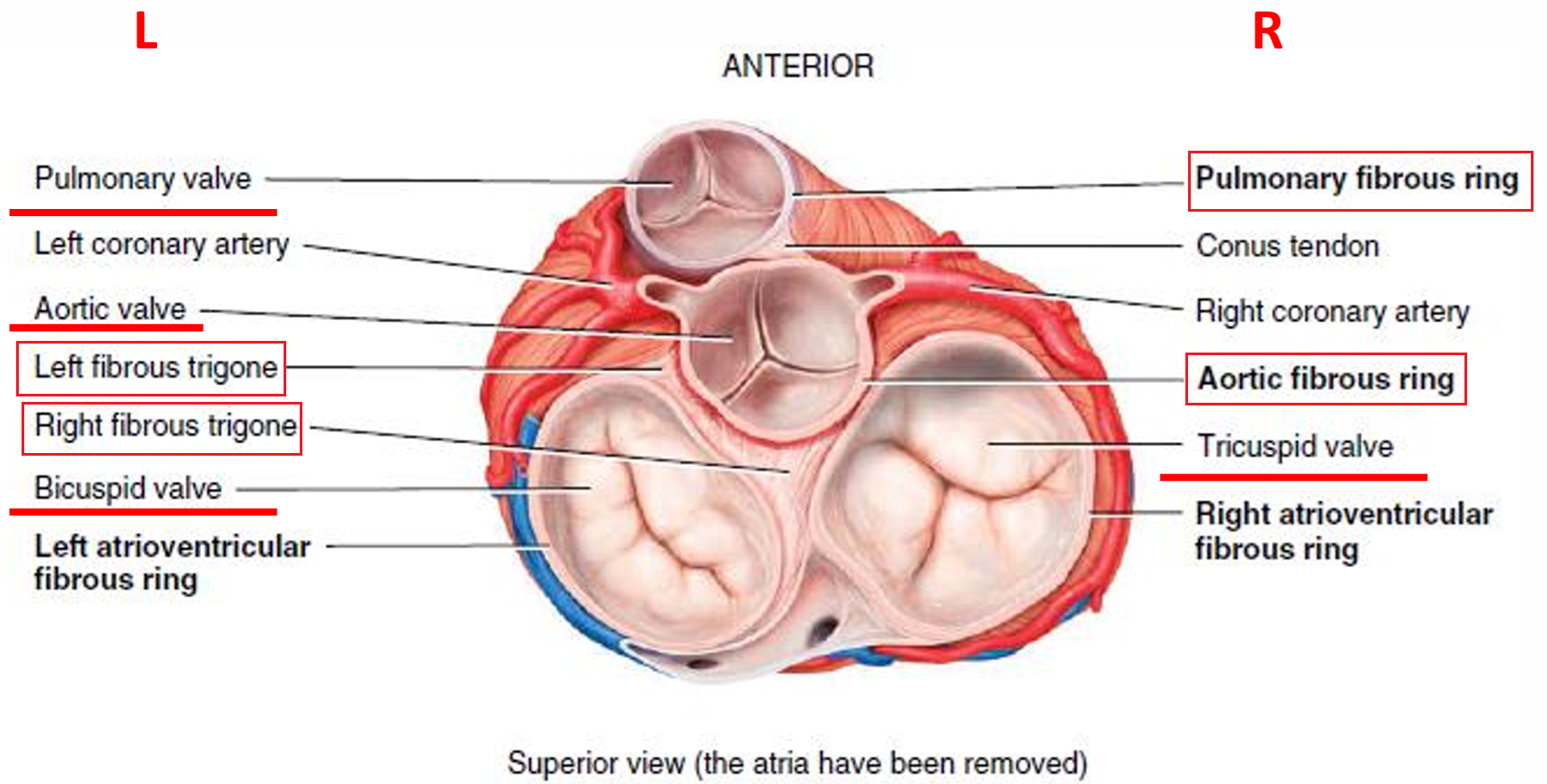
- Four dense connective tissue rings surround the valves of the heart
- form a structural foundation for the heart valves
- prevent overstretching of the valves as blood passes through them
- acts as an electrical insulator between the atria and ventricles
- 图
- Location
- conduction sysytem
- Autorhythmic fibers - specialized cardiac muscle fibers
- Self-excitable - act as a pacemaker, generate AP
- Cardiac chambers contract in a coordinated manner
- 兴奋过程
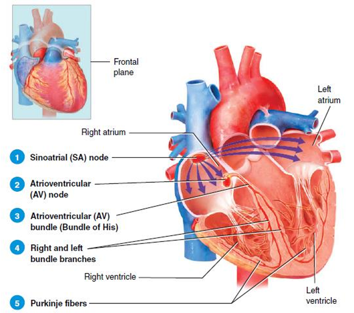
- Cardiac excitation begins in the sinoatrial node
- AP propagates through both atria via intercalated discs
- Both atria contract
- AP reaches the atrioventricular node
- AP enters the atrioventricular bundle 房室束(希氏束)
- AP enters both the right and left bundle branches 右束支和左束支
- AP propagates along Purkinje fibers
- Both ventricles contract
- 图
- Coronary Circulation
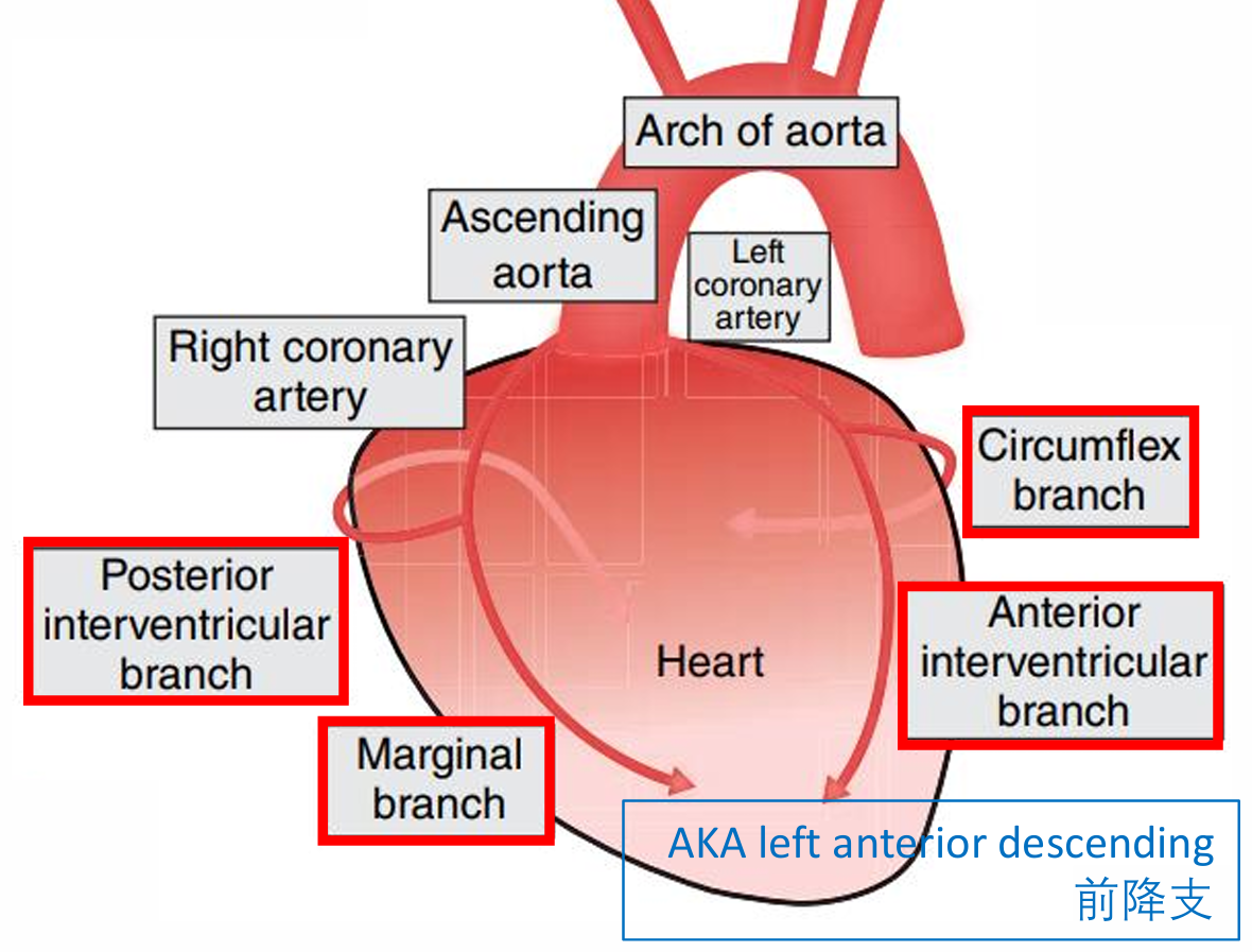
- Coronary arteries
- branch from the ascending aorta
- supply oxygenated blood to the myocardium
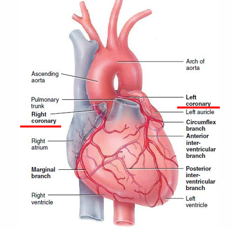
- left coronary artery - passes inferior to the left auricle
- right coronary artery - passes inferior to the right auricle
- 图
- 图
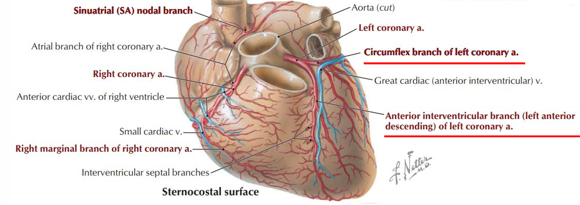
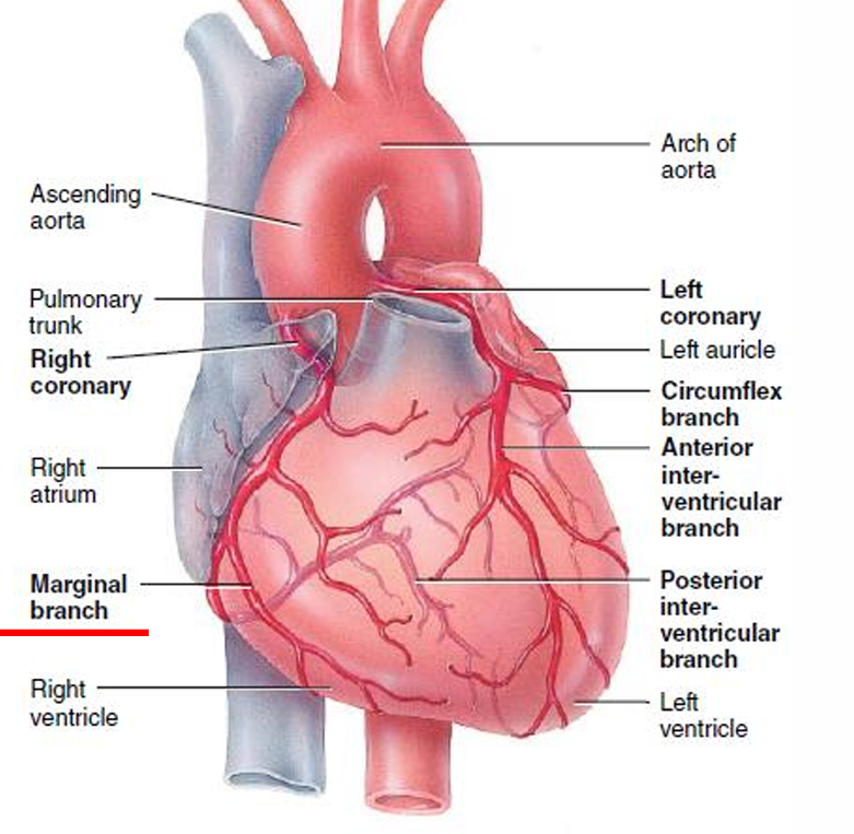
- Left coronary artery
- passes inferior to the left auricle and divides into
- Left anterior descending (LAD)
- in the anterior interventricular sulcus 走行于前室间沟内
- supplies both ventricles
- Circumflex branch 旋支
- in the coronary sulcus 走行于冠状沟内
- supplies both atria
- Left anterior descending (LAD)
- 图
- passes inferior to the left auricle and divides into
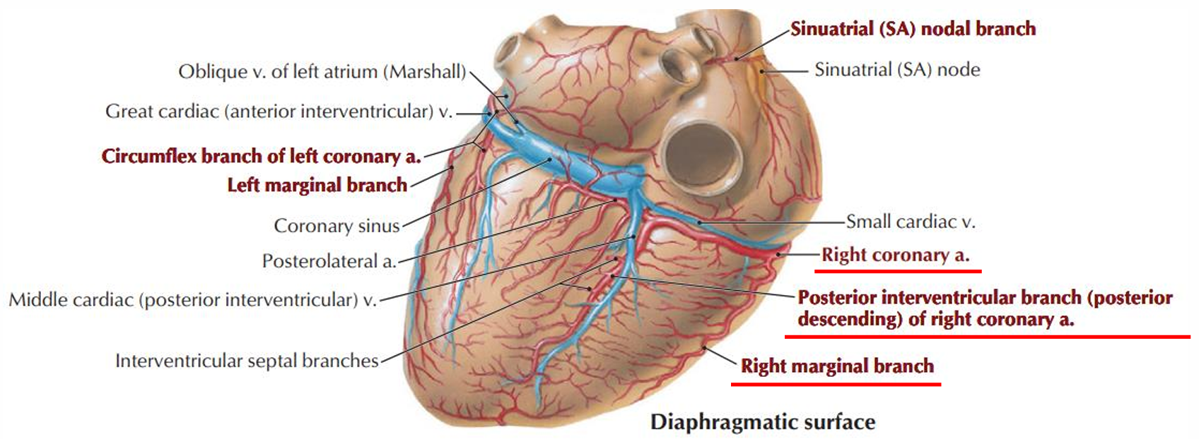
- Right coronary artery
- passes inferior to the right auricle and divides into
- marginal branch 右缘支
- runs along the right margin of the heart 走行于心脏右缘
- supplies RV
- 图
- posterior descending of right coronary A 后降支
- in the posterior interventricular sulcus 走行于后室间沟
- supplies both ventricles
- 图
- marginal branch 右缘支
- passes inferior to the right auricle and divides into
- Coronary arteries
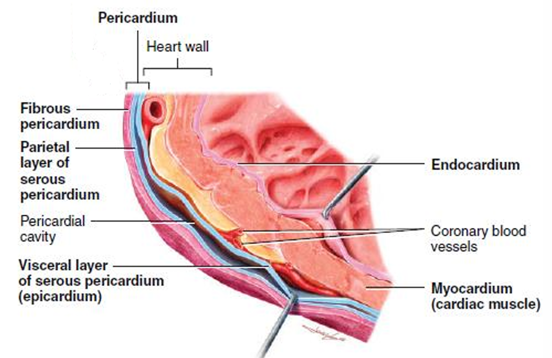
- Pericardium
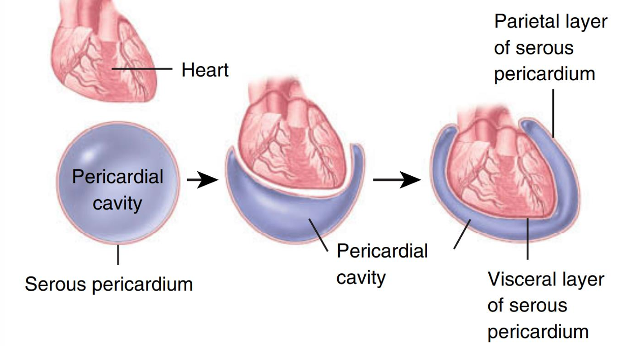
- Pericardium is triple-layered membrane that surrounds the heart
- 分为
- the fibrous pericardium 纤维性心包
- 在浆膜性心包外面
- 在浆膜性心包外面
- the serous pericardium 浆膜性心包
- 2层
- parietal layer
- visceral layer, AKA epicardium 心外膜
- pericardial cavity - cavity b/t the parietal and visceral layers 心包腔
- pericardial fluid - lubricating serous fluid 心包液
- 图
- 2层
- the fibrous pericardium 纤维性心包
- Heart Wall
- Epicardium (external layer)
- Myocardium (middle layer)
- composed of cardiac muscle tissue
- makes up approximately 95% of the heart wall
- responsible for the pumping action
- Endocardium (inner layer)
- thin layer of connective tissue
- minimizes the surface friction as blood passes through the heart
- Aorta
- Ascending aorta
- locates posterior to the pulmonary trunk
- gives off 2 coronary arteries
- arches to the left
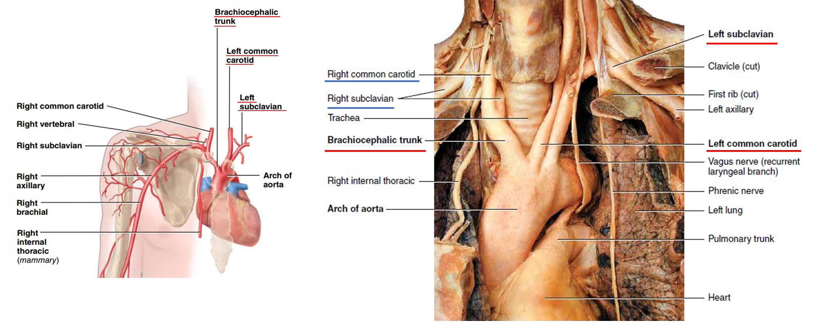
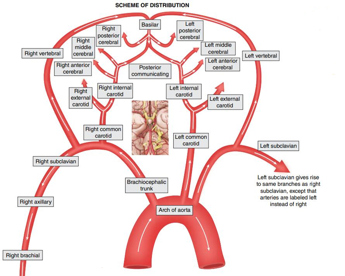
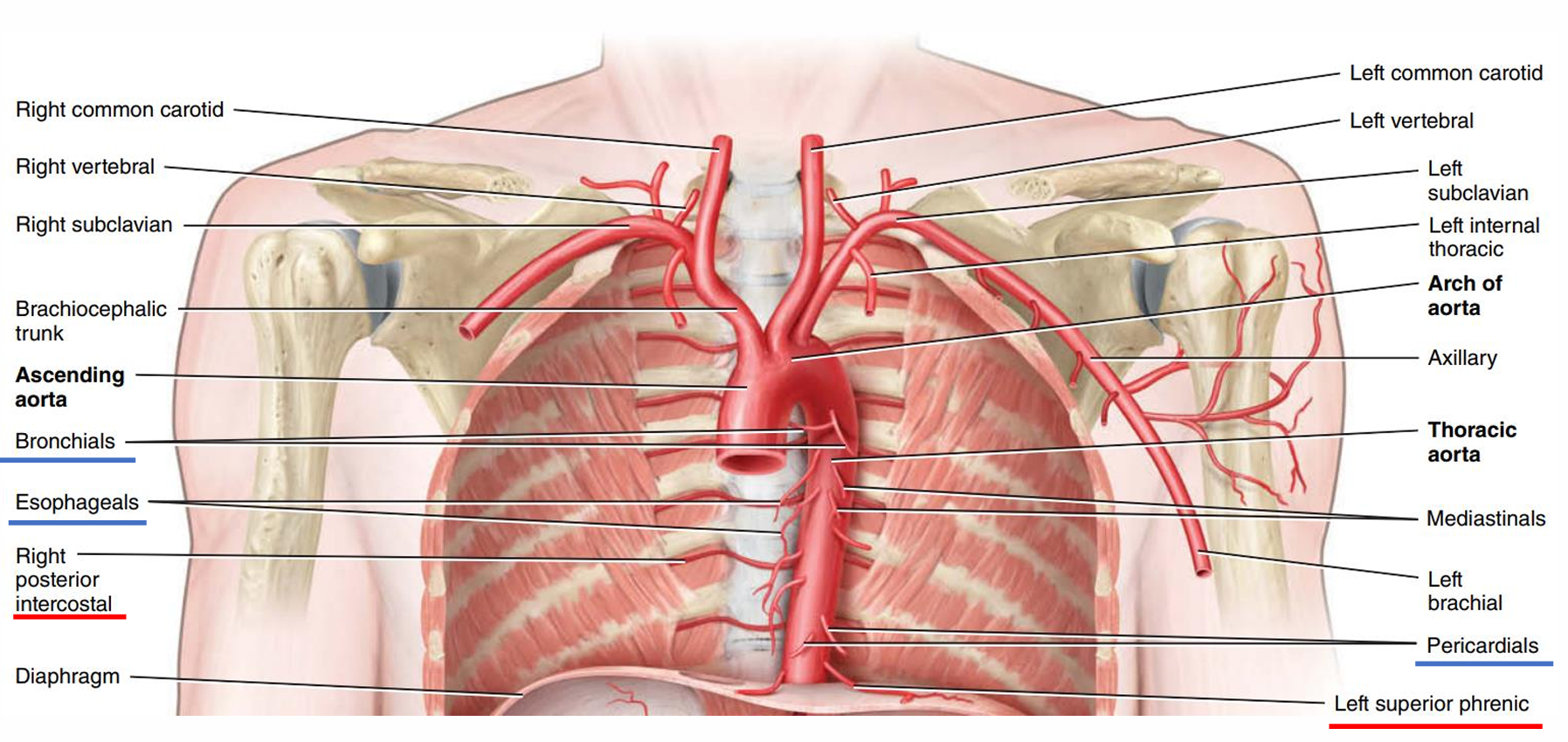
- Arch of the aorta
- 左右在刚开始不相同分支
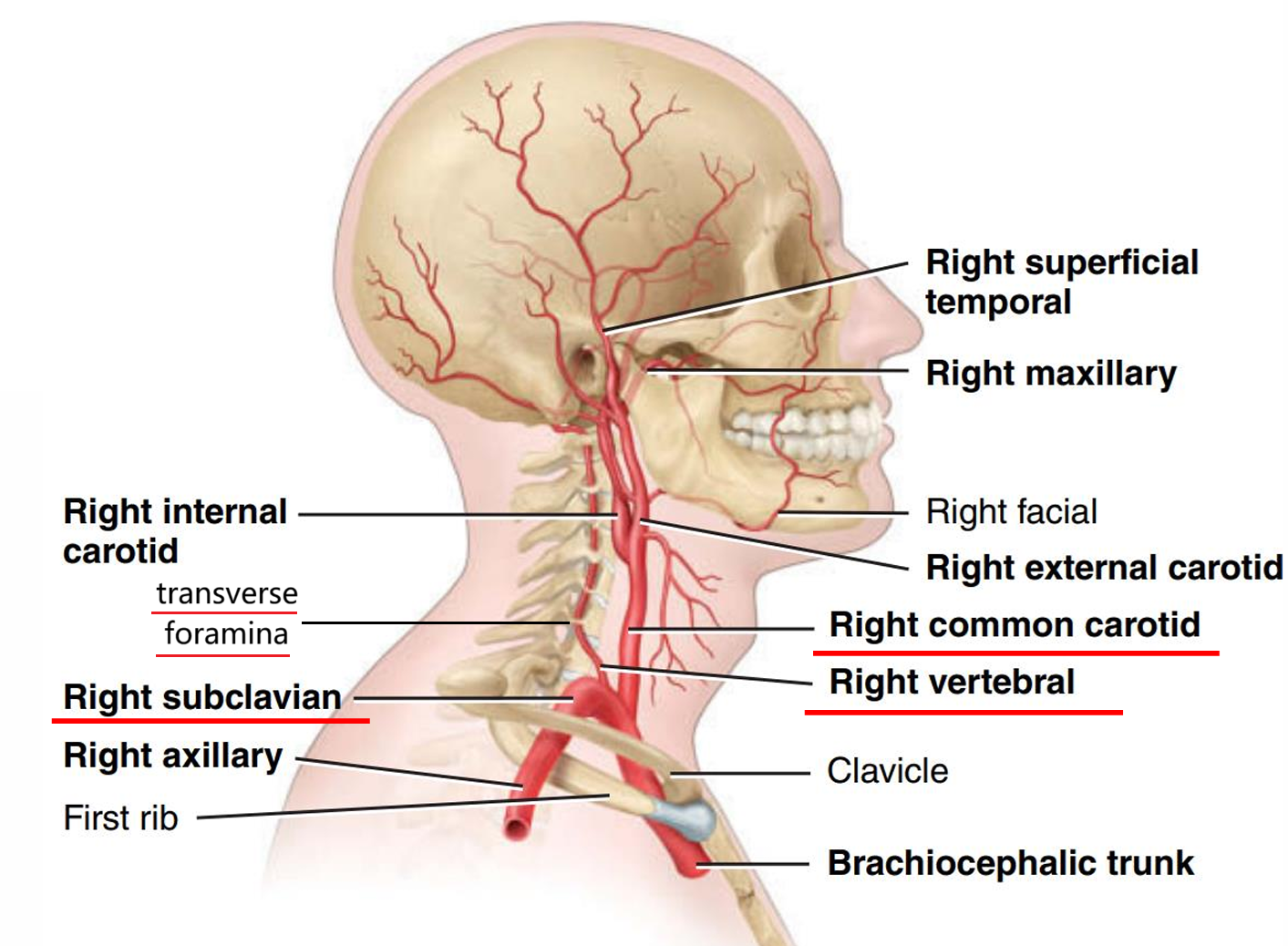
- brachiocephalic trunk
- right common carotid A
- right subclavian A
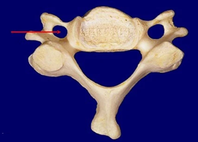
- right vertebral A
- 横突孔在颈椎上
- 横突孔在颈椎上
- 图
- left common carotid
- left subclavian
- left vertebral
- 先分成三个分支
- 图
- brachiocephalic trunk
- 在后面左右对称
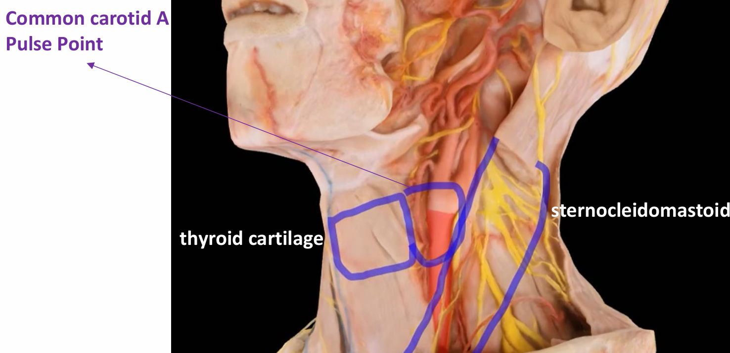
- Common carotid A
- Ascending aorta → brachiocephalic trunk → right common carotid
- Ascending aorta → left common carotid
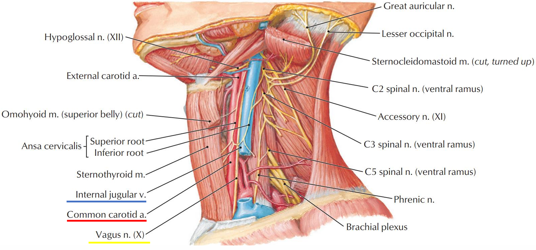
- Carotid sheath颈动脉鞘 contains the common carotid A, internal jugular V, and vagus N.
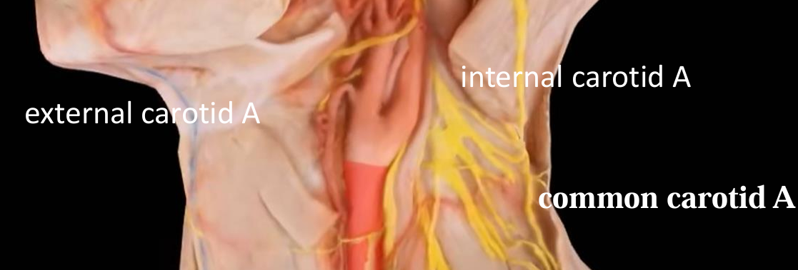
- common carotid A在颈动脉窦部分分成internal carotid A与external carotid A
- pressure-monitoring receptors in the carotid sinus 颈动脉窦
- chemoreceptors (receptors that monitor blood levels of oxygen and carbon dioxide) in the carotid bodies 颈动脉小球
- 分叉的点在喉结高度的地方
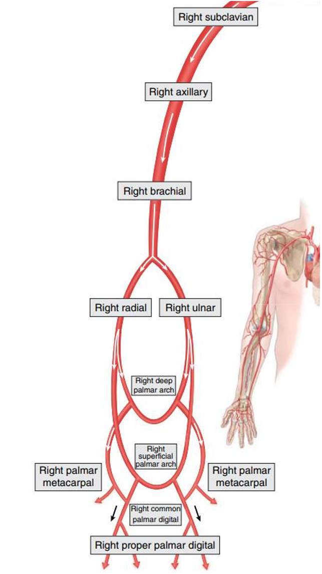
- Arteries of the upper limbs
- Subclavian A
- Axillary A 腋动脉
- Brachial A 肱动脉
- Radial A 桡动脉
- Ulnar A 尺动脉
- Deep palmar arch 掌深弓
- Superficial palmar arch 掌浅弓
- 左右对称
- Common carotid A
- 左右在刚开始不相同分支
- Descending aorta
- Thoracic aorta
- Parietal branches 壁支
- Posterior intercostal A (9 pairs) 肋间后动脉
- arise from posterolateral aspect of thoracic aorta
- Start inferior to 3rd rib
- run through intercostal space
- supply intercostal and chest muscles
- Subcostal A (1 pair) 肋下动脉
- inferior to 12th rib
- supply upper abdominal muscles
- Superior phrenic A (1 pair) 膈上动脉
- supply diaphragm
- Posterior intercostal A (9 pairs) 肋间后动脉
- Visceral branches 脏支
- Bronchials 支气管支
- Esophageals 食管支
- Pericardials 心包支
- 图
- Parietal branches 壁支
- Abdominal aorta
- Unpaired visceral branches
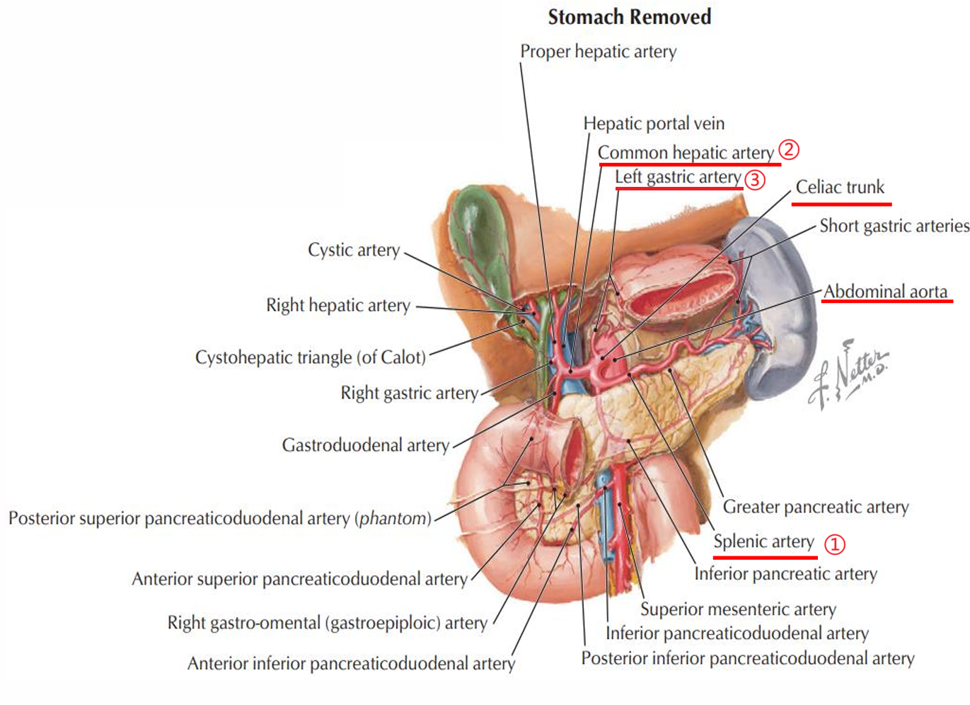
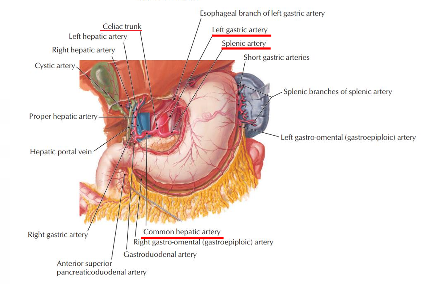
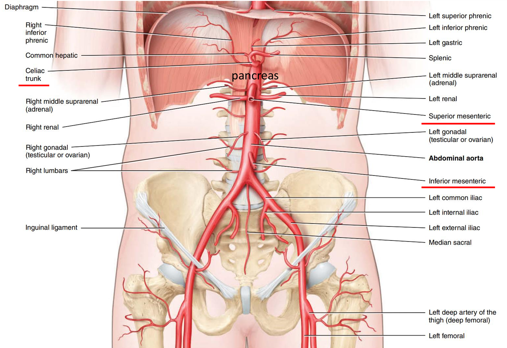
- Celiac trunk 腹腔干
- Splenic artery
- Common hepatic artery
- Left gastric artery
- 没胃
- 有胃
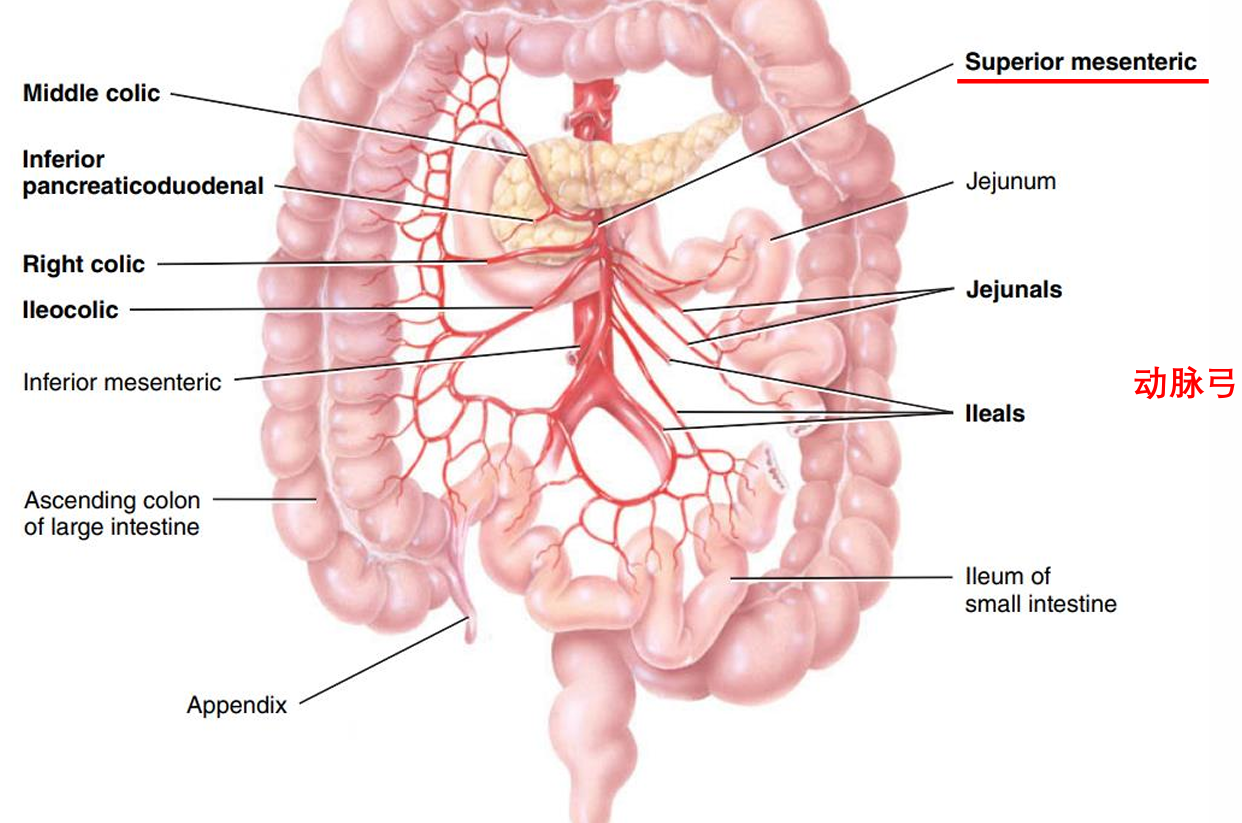
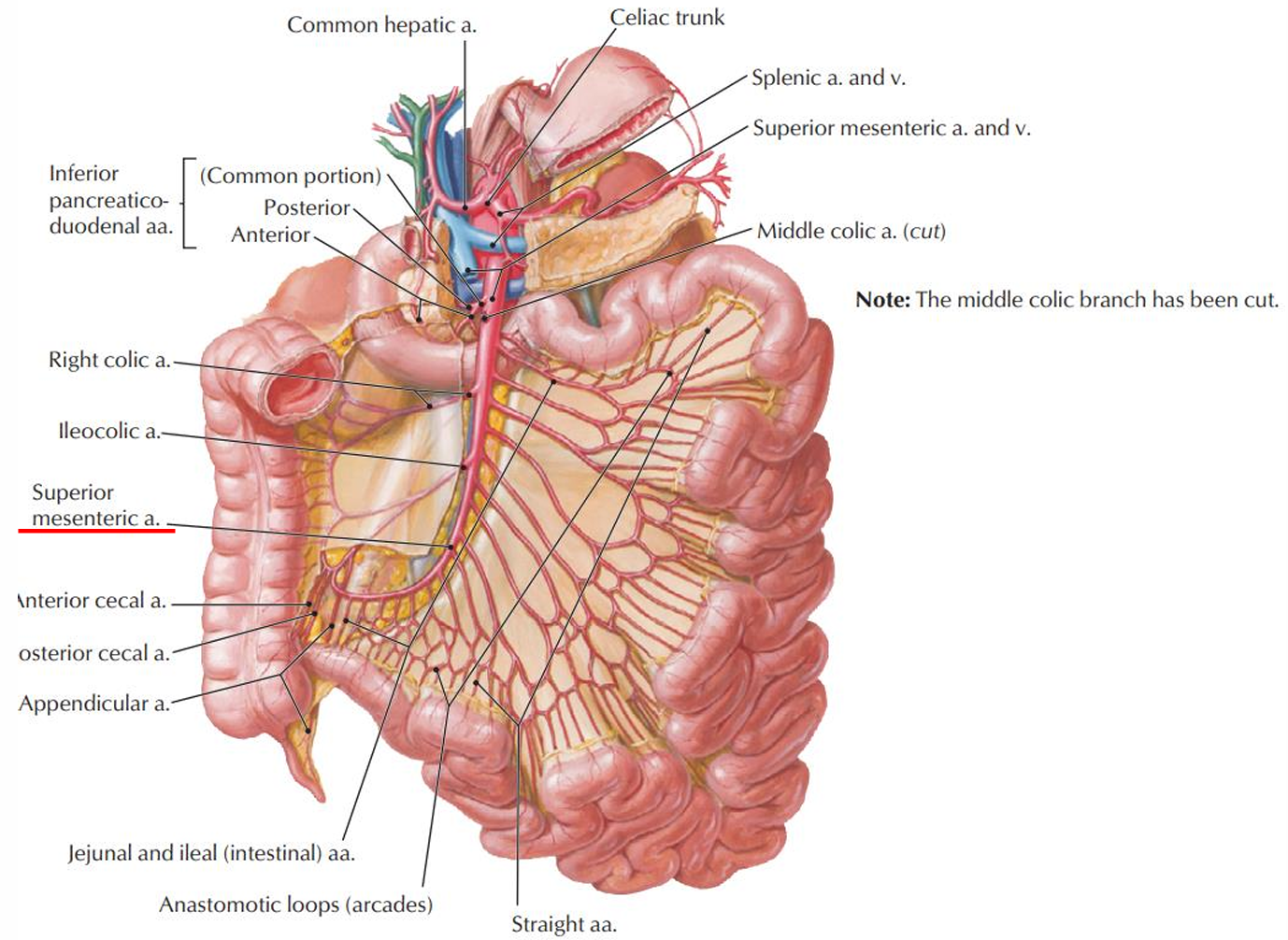
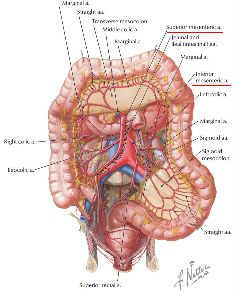
- Superior mesenteric A 肠系膜上动脉
- between pancreas and duodenum
- supplies from duodenum to transverse colon
- between pancreas and duodenum
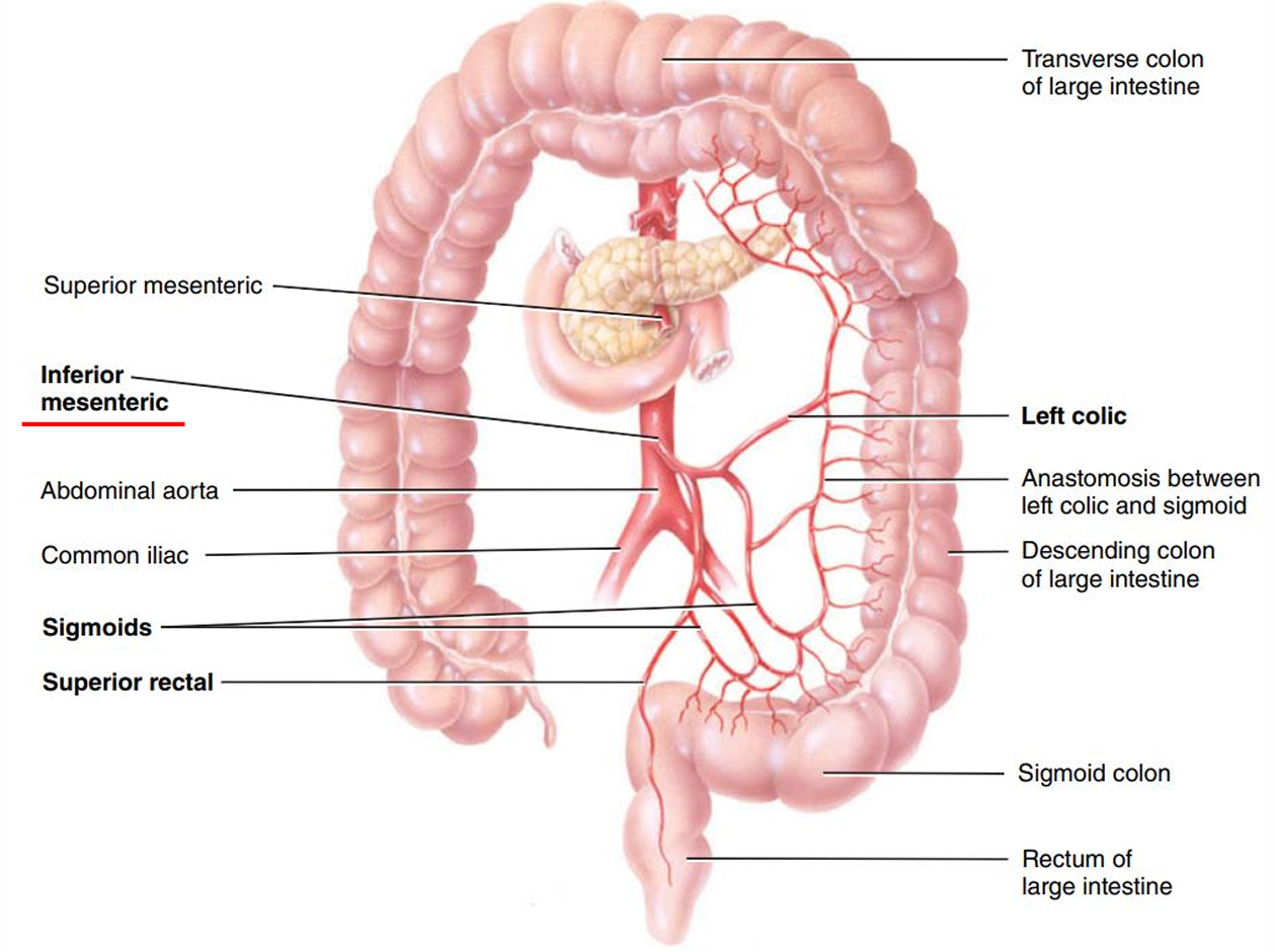
- Inferior mesenteric A 肠系膜下动脉
- Inferior to duodenum
- supplies from transverse colon to rectum
- Inferior to duodenum
- 图
- Celiac trunk 腹腔干
- Paired visceral branches
- Middle suprarenal A 肾上腺中动脉
- Renal A 肾动脉
- Testicular A/Ovarian A 睾丸、卵巢动脉
- Unpaired parietal branch
- Median sacral A 骶正中动脉
- Paired parietal branch
- Inferior phrenic A (1 pair) 膈下动脉
- Lumbar A (4 pairs) 腰动脉
- Unpaired visceral branches
- Arteries of the pelvis and lower limbs
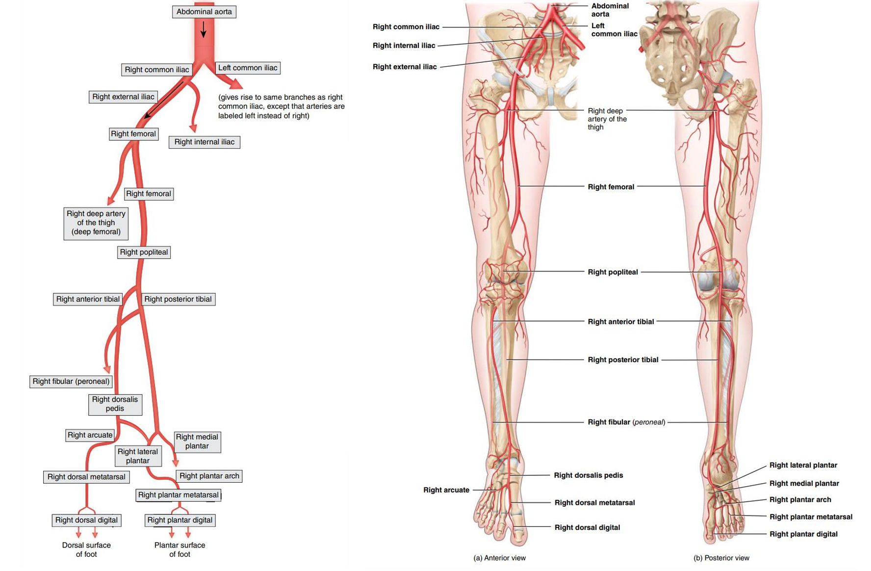
- right and left common iliac A
- internal iliac A
- external iliac A
- femoral A
- popliteal A 腘动脉
- anterior and posterior tibial A 胫前动脉和胫后动脉
- 图
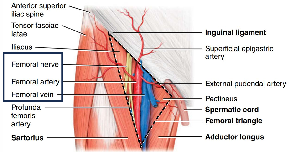
- Femoral triangle
- SAIL - Sartorius + Adductor longus 长收肌+ Inguinal Ligament 腹股沟韧带
- 这个区域有动脉, 静脉, 神经
- SAIL - Sartorius + Adductor longus 长收肌+ Inguinal Ligament 腹股沟韧带
- right and left common iliac A
- Thoracic aorta
- Ascending aorta
- Pulmonary circulation
- deoxygenated blood
- RV → pulmonary trunk →
- right pulmonary artery → right lung
- left pulmonary artery → left lung
- RV → pulmonary trunk →
- oxygenated blood
- ① Left upper lobe
- ② Left lower lobe
- ③ Right upper and middle lobe
- ④ Right lower lobe
- 图
- deoxygenated blood
- Veins
- 基础
- Venous valves
- Valve cusps point toward heart
- allow one-way flow toward the heart
- Prevent the backflow of blood when muscles relax
- Valve cusps point toward heart
- Mechanisms of venous return
- Venous return - the flow of blood from the periphery back to the RA
- Mechanisms
- Pressure gradient
- Skeletal muscle pump
- e.g. contraction of gastrocnemius腓肠肌 prompts venous return
- Artery vs. Vein
- 单词的区别
- Artery 叫 Branch
- Vein 叫 Tributary - smaller veins draining into larger veins
- Carotid 颈动脉的
- Common carotid A
- External carotid A
- Internal carotid A
- Jugular 颈静脉的
- External jugular V
- Internal jugular V
- arteries are deep, veins may be superficial or deep, deep veins go alongside arteries
- 单词的区别
- Venous valves
- three systemic veins into RA
- coronary sinus
- superior vena cava
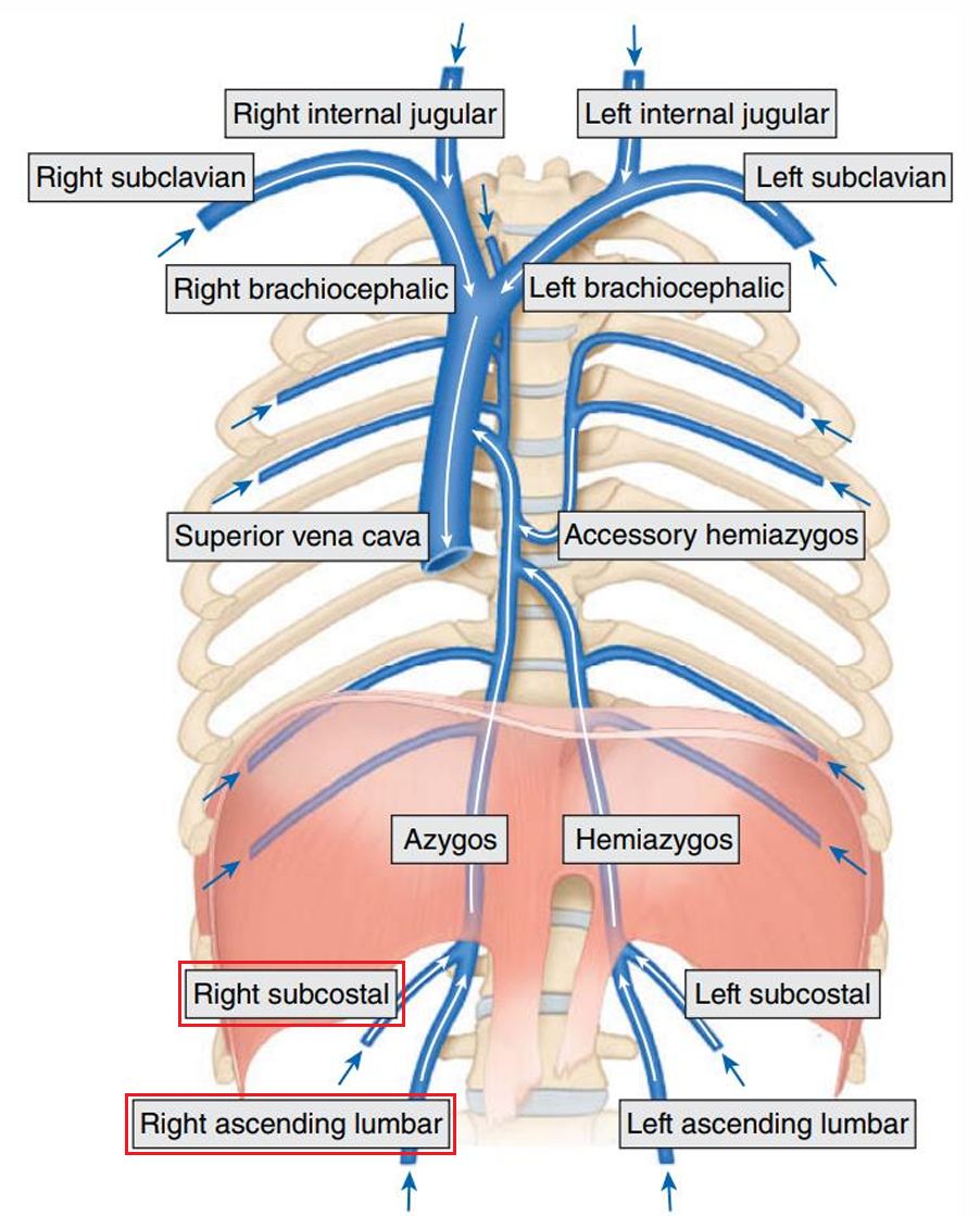
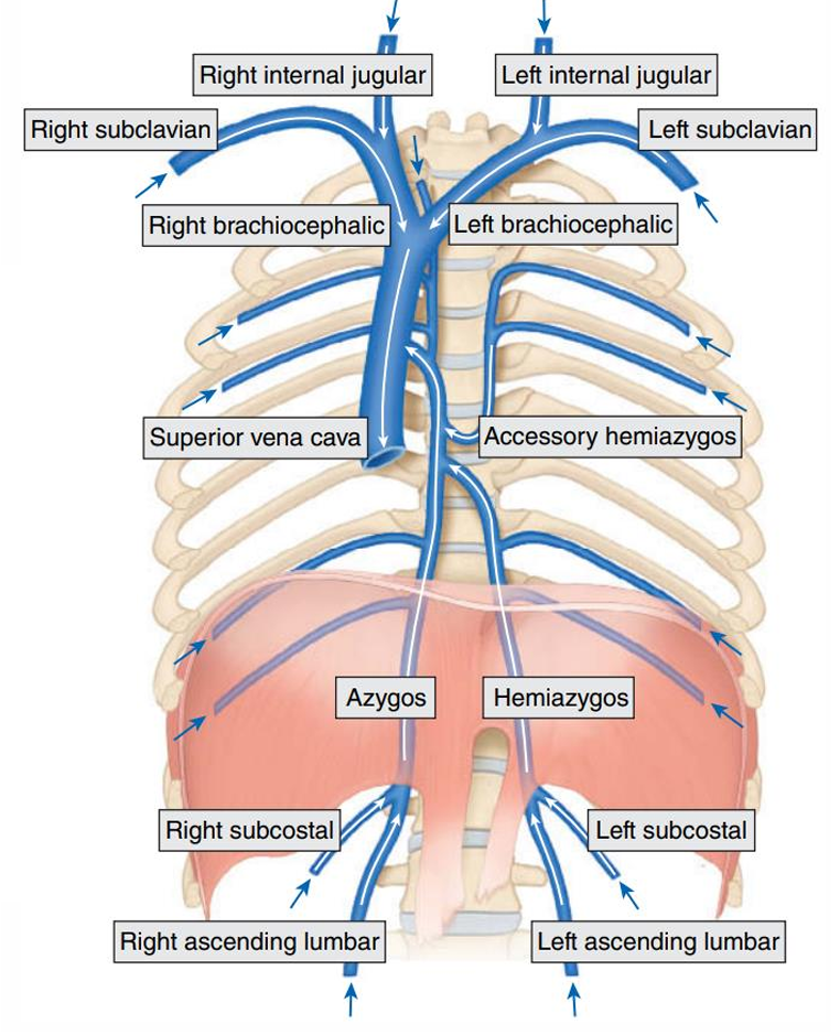
- Azygos system
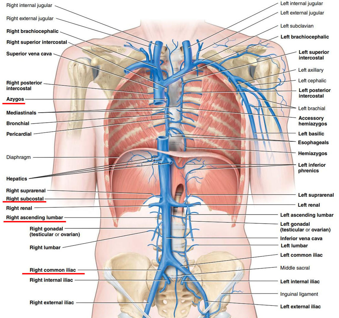
- Azygos V
- only on the right side of the vertebral column
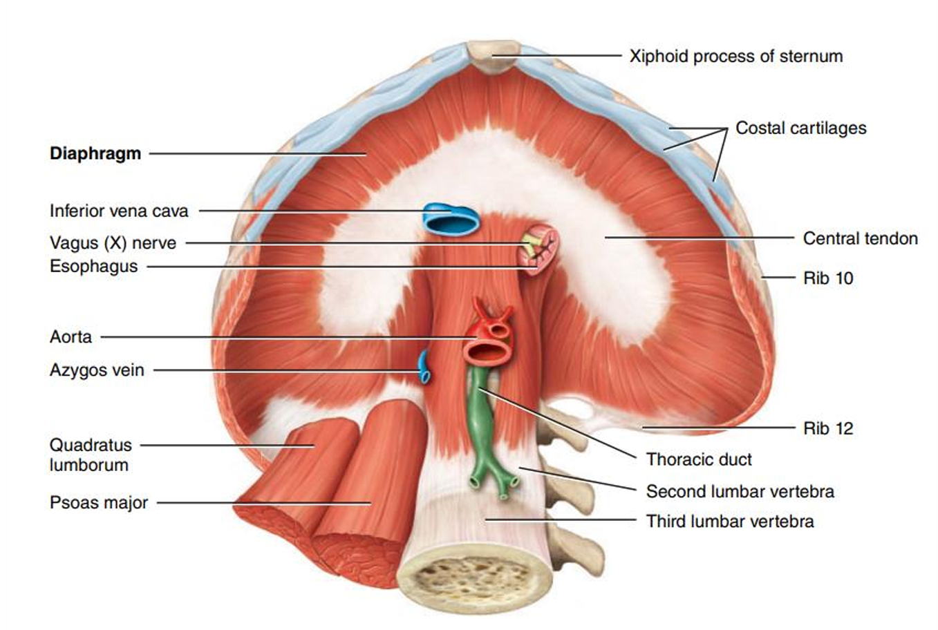
- begins at junction of right ascending lumbar V右腰升静脉 and right subcostal V右肋下静脉 near diaphragm
- right ascending lumber V 右腰升静脉 is a tributary of common iliac V
- 穿过diaphragm
- Hemiazygos V
- Accessory hemiazygos V
- Azygos V
- Brachiocephalic veins
- 分为 Right Brachiocephalic veins(3cm) 与 Left Brachiocephalic veins(6cm)
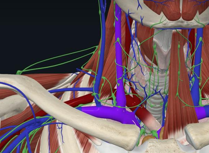
- Subclavian V + Internal jugular V → brachiocephalic V → superior vena cava 都由两个静脉汇入
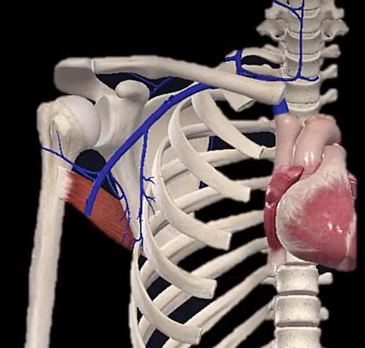
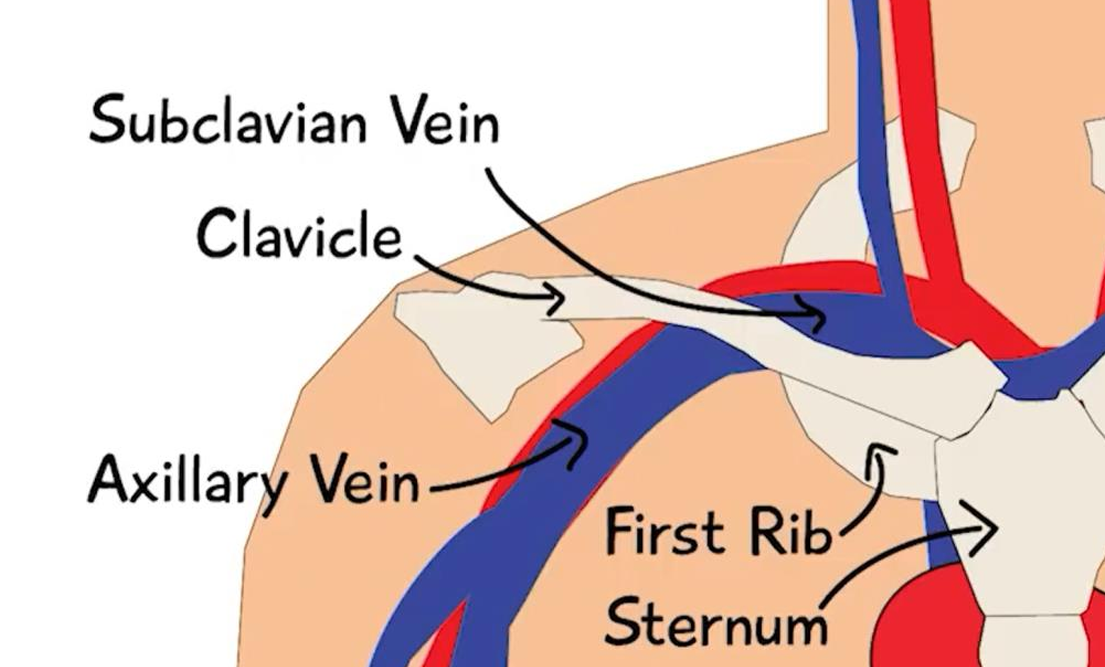
- Subclavian V
- 位置
- inferior to minor pectoral muscle
- inferior to clavicle
- superior to 1st rib
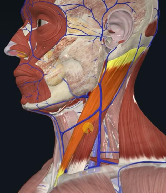
- External jugular veins 颈外静脉
- Across sternocleidomastoid 胸锁乳突肌
- External jugular vein → subclavian V
- Become very prominent凸显 along side of neck when venous pressure rises
- Across sternocleidomastoid 胸锁乳突肌
- Axillary vein 腋静脉
- 位置
- Axillary vein → Subclavian V
- Starts at border of the teres major 大圆肌边缘
- Ends at subclavian V
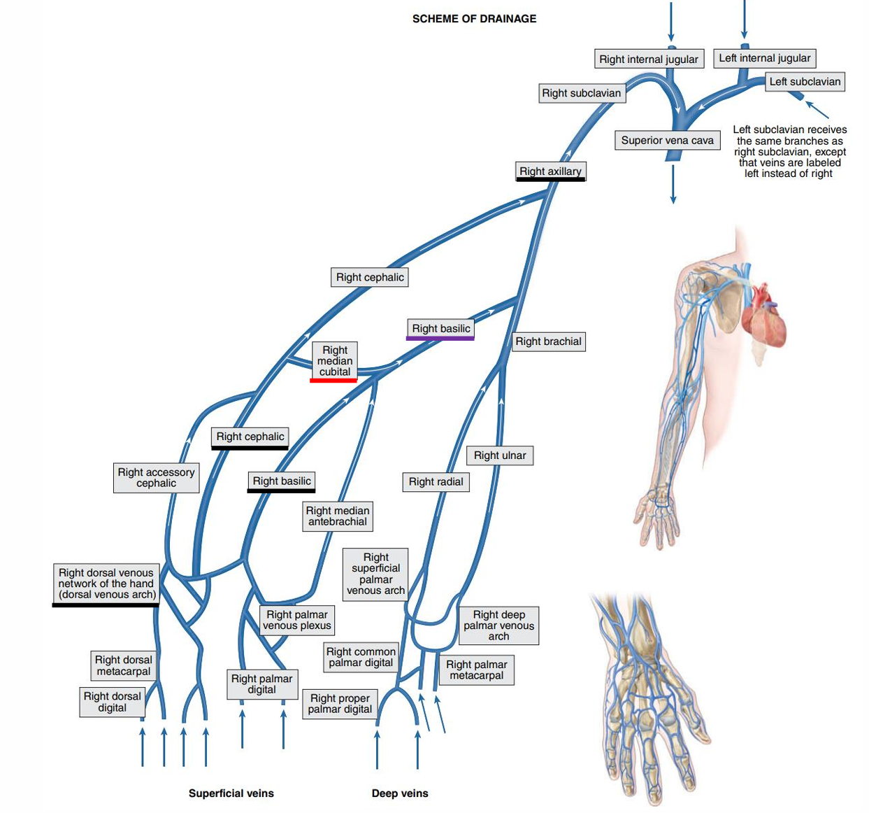
- Veins of the Upper Limbs
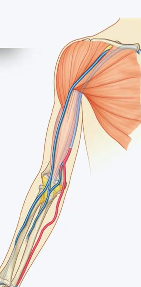
- Deep veins
- 特点
- Locate deep in the body
- Follow the course of the arteries
- Two veins accompany one artery
- Have the same names as the corresponding arteries
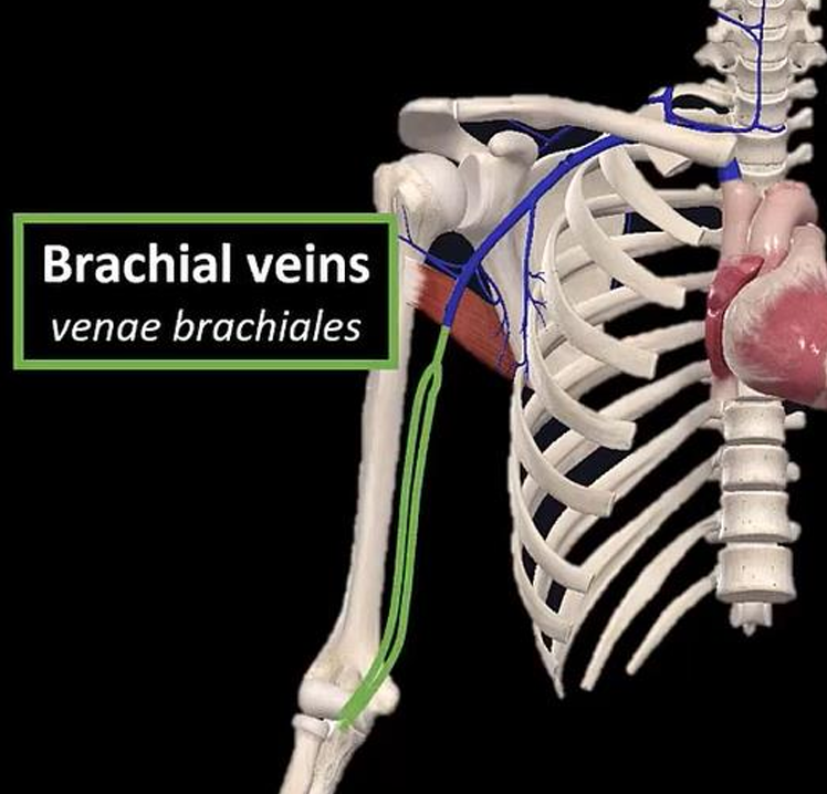
- Brachial veins 肱静脉
- Two brachial veins
- 起点 - cubital fossa
- 终点 - distal border of teres major m
- 图
- Ulnar veins
- Radial veins
- Deep venous palmar arch
- 特点
- Superficial veins
- 特点
- locate just deep to the skin and are often visible
- do not accompany arteries
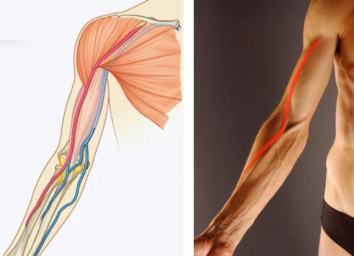
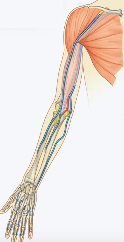
- Basilic vein 贵要静脉
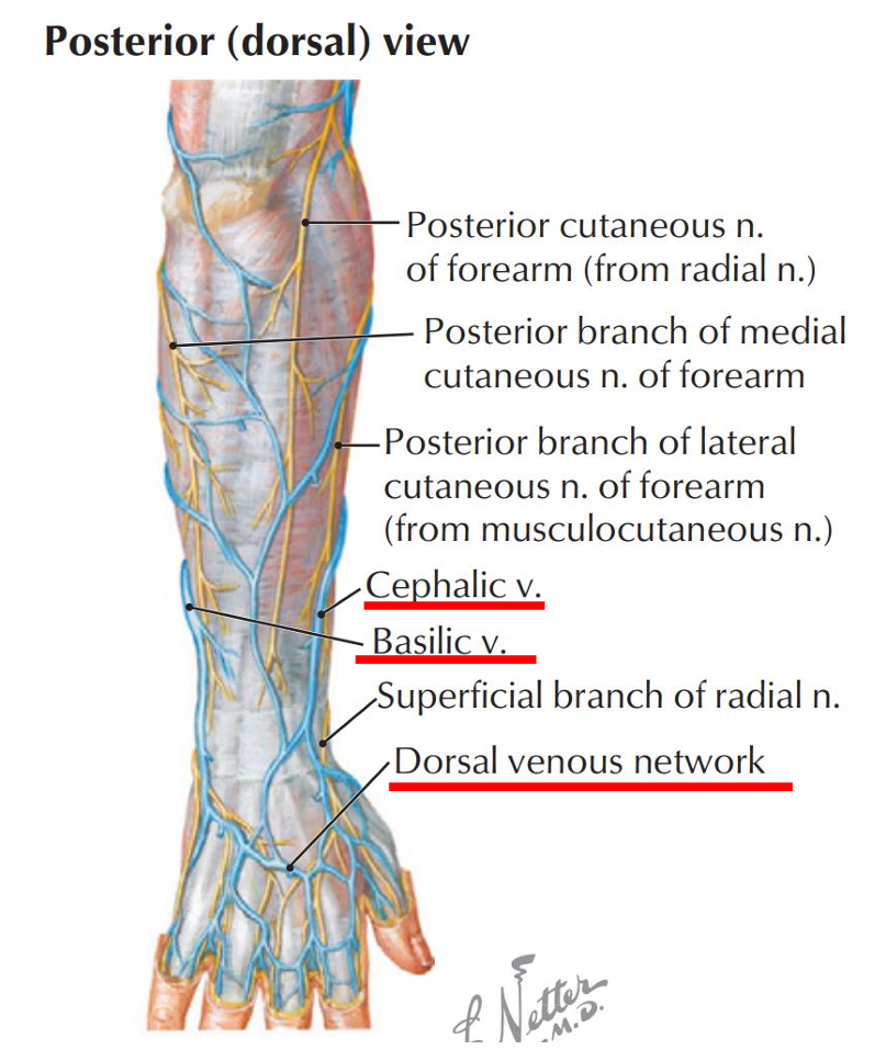
- Starts at the dorsal venous network
- Courses on the medial side of the forearm
- Basilic vein → the brachial V → Axillary vein
- 红色这段
- Cephalic vein 头静脉
- Starts at the dorsal venous network
- Courses on the lateral side of the forearm
- Cephalic vein → Axillary V
- 红色这段
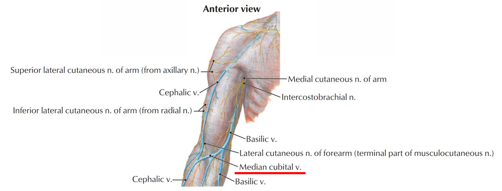
- Median cubital vein
- Connects cephalic V and basilic V in cubital fossa 肘窝
- Commonly used for venipuncture 静脉穿刺
- Connects cephalic V and basilic V in cubital fossa 肘窝
- Intercaptitular vein
- Dorsal venous network of hand
- Venous network on the dorsum of the hand
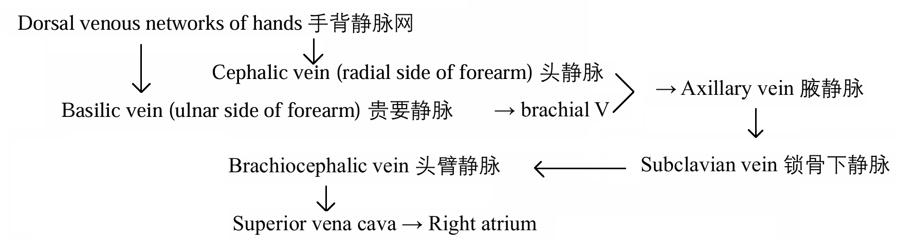
- lateral aspect of dorsal venous network → Cephalic V (radial side of forearm) 头静脉
- medial aspect of dorsal venous network → Basilic V (ulnar side of forearm) 贵要静脉
- 图
- 特点
- Deep veins
- 位置
- 位置
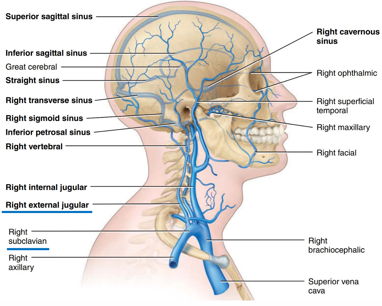
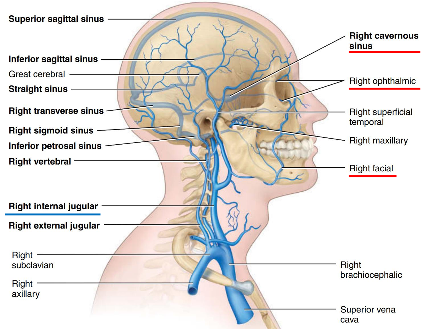
- Internal jugular V
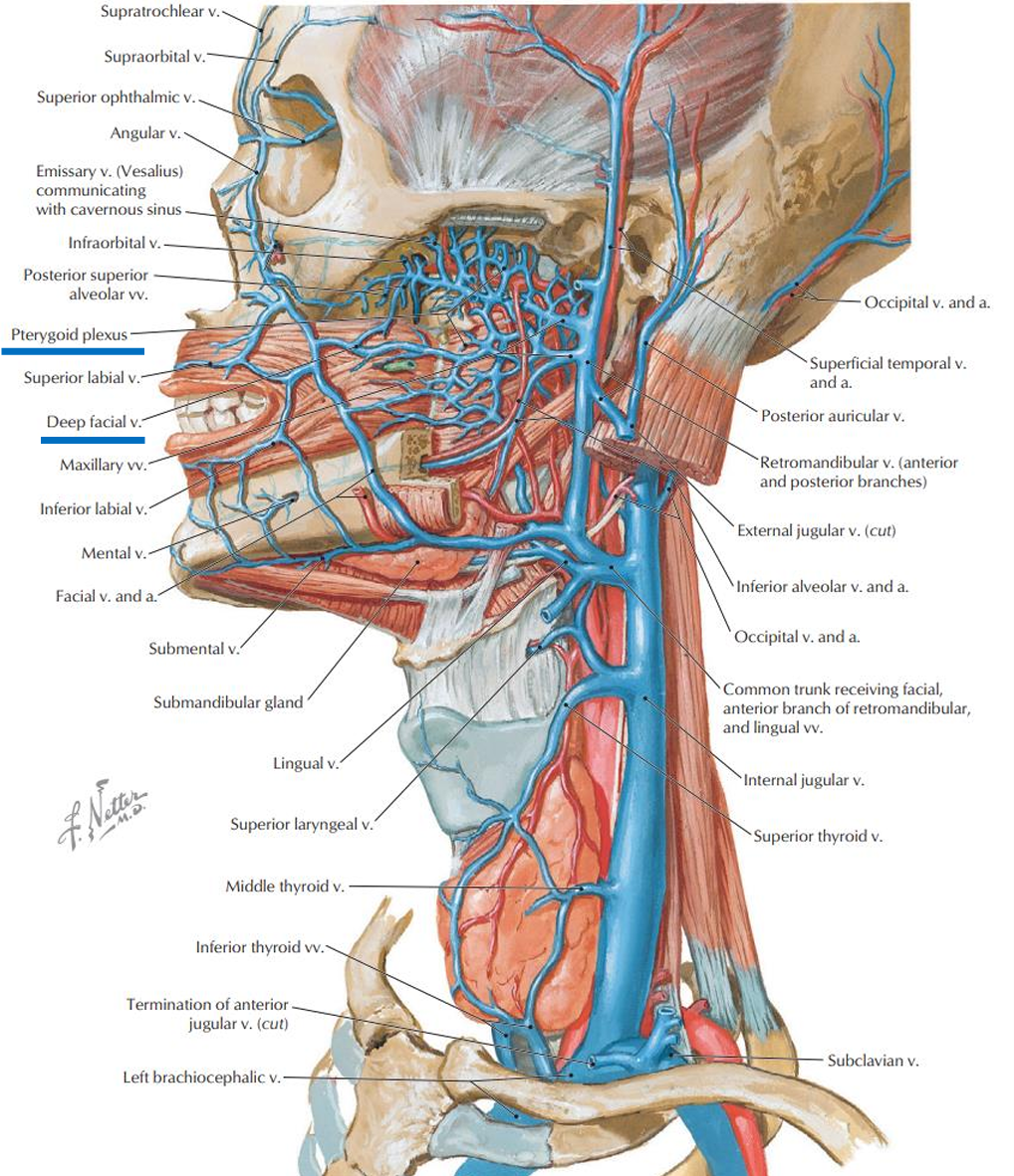
- facial vein
- Ophthalmic V 眼静脉
- 路径
- 图
- 路径
- Deep facial V
- Deep facial V → Pterygoid plexus 翼静脉丛 → Cavernous sinus
- Deep facial V → Pterygoid plexus 翼静脉丛 → Cavernous sinus
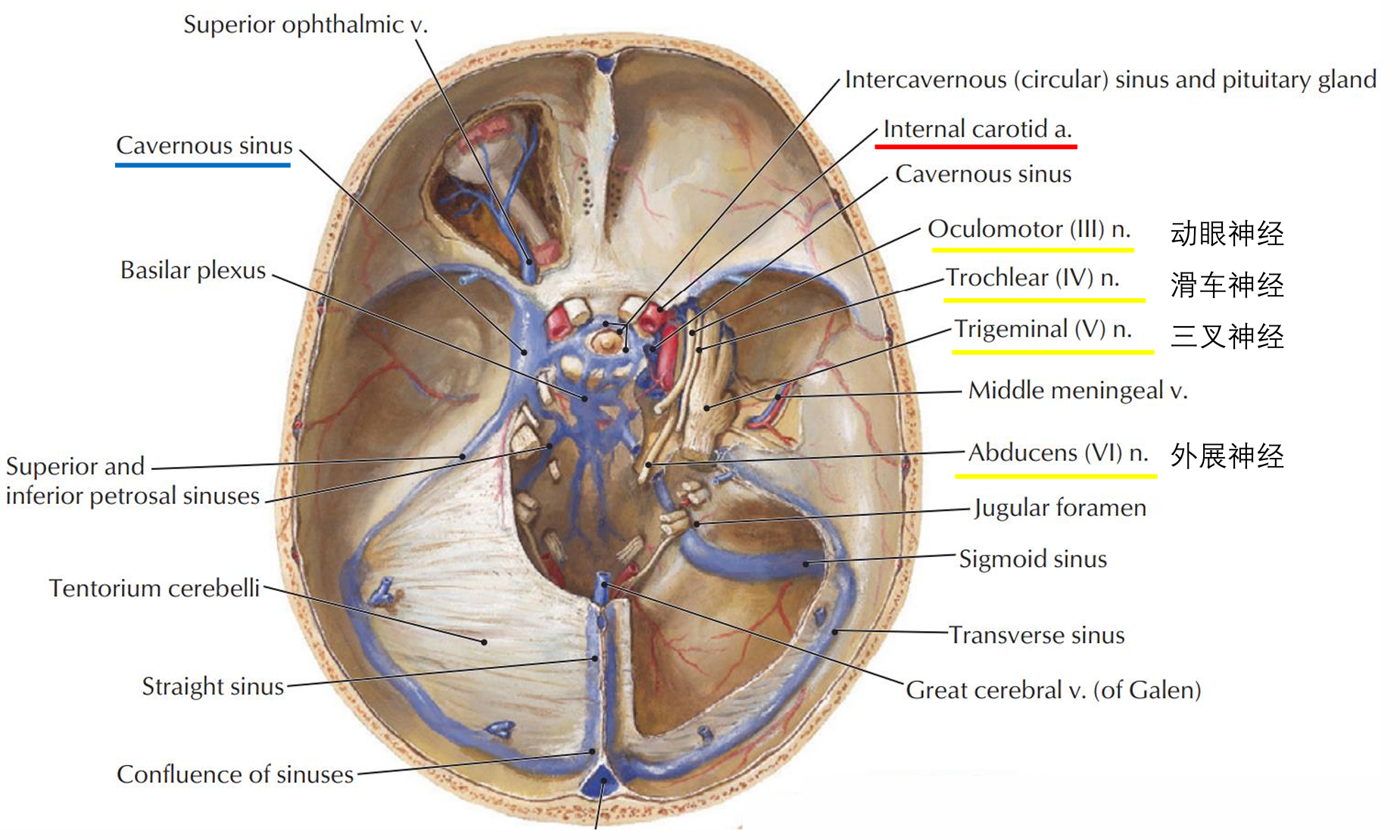
- Cavernous sinus 海绵窦
- 连着许多静脉,动脉,神经感染会有危险
- 连着许多静脉,动脉,神经感染会有危险
- Ophthalmic V 眼静脉
- 病-CVC
- Central Venous Catheter 中心静脉导管
- IJV是CVC最常见的穿刺部位之一
- facial vein
- 总结
- 从手背静脉注射的路径
- 图
- 从手背静脉注射的路径
- Azygos system
- inferior vena cava
- 基础
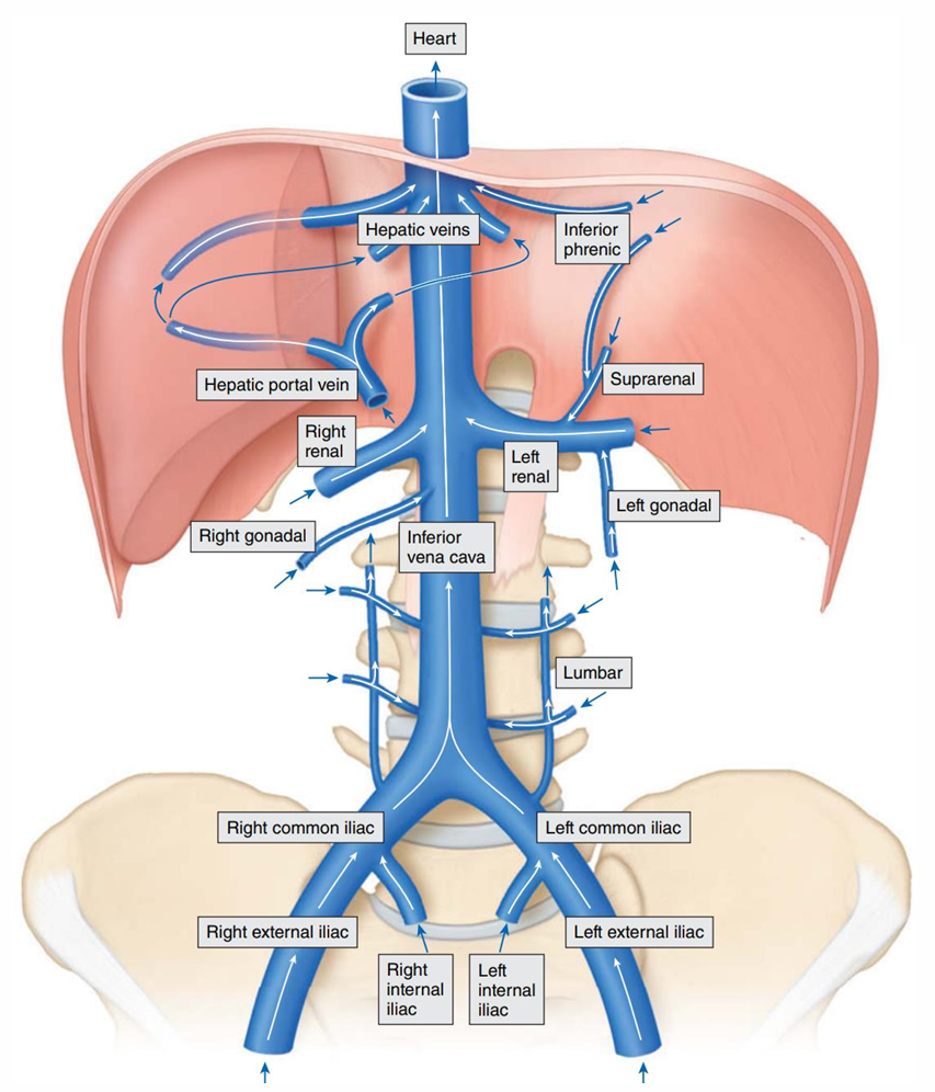
- Blood from abdominal and pelvic viscera returns to inferior vena cava. 下腔静脉的属支
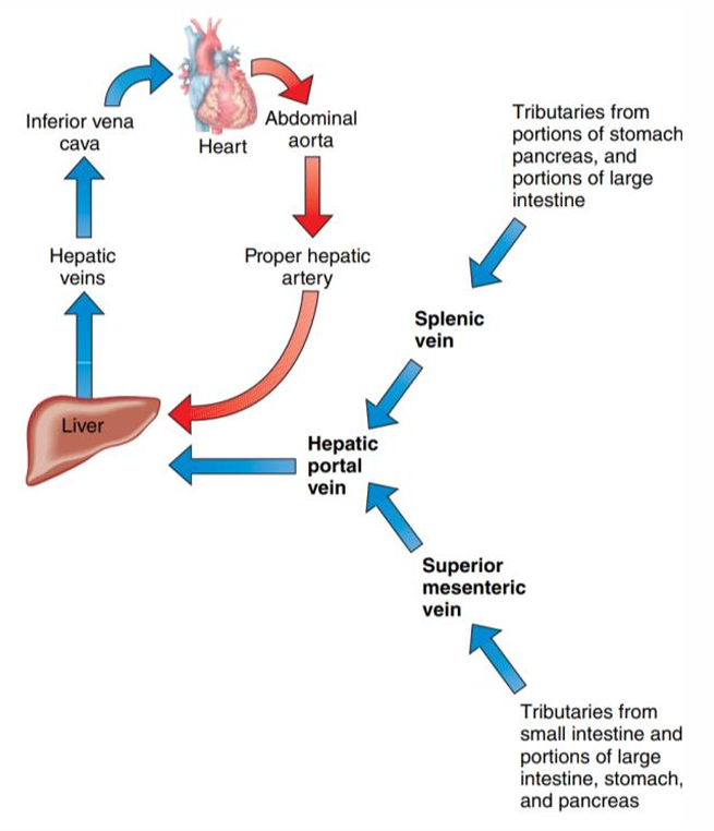
- Bood from GI tract, spleen, pancreas, and gallbladder return to hepatic portal vein. 肝门静脉系
- Collects blood from the lower half of the body (below the diaphragm)
- No valves
- Starts at the junction of the right and left common iliac V
- Runs on the right side of the vertebral column
- Runs on the right of the abdominal aorta
- Visceral tributaries
- Hepatic V
- Left renal V
- Left suprarenal V
- Left ovarian/testicular V
- Right renal V
- Right suprarenal V
- Right ovarian/testicular V
- Parietal tributaries
- Lumbar V (4 pairs)
- Inferior phrenic V
- Veins of the lower limbs
- 分类
- Superficial veins
- In subcutaneous layer
- Anastomose with one another and with deep veins
- Do not go with arteries
- Deep veins
- Go with arteries
- Have the same names as corresponding arteries
- All veins of the lower limbs have valves, which are more numerous than in veins of the upper limbs.
- Superficial veins
- Veins of thigh
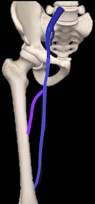
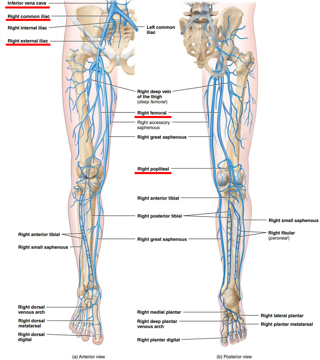
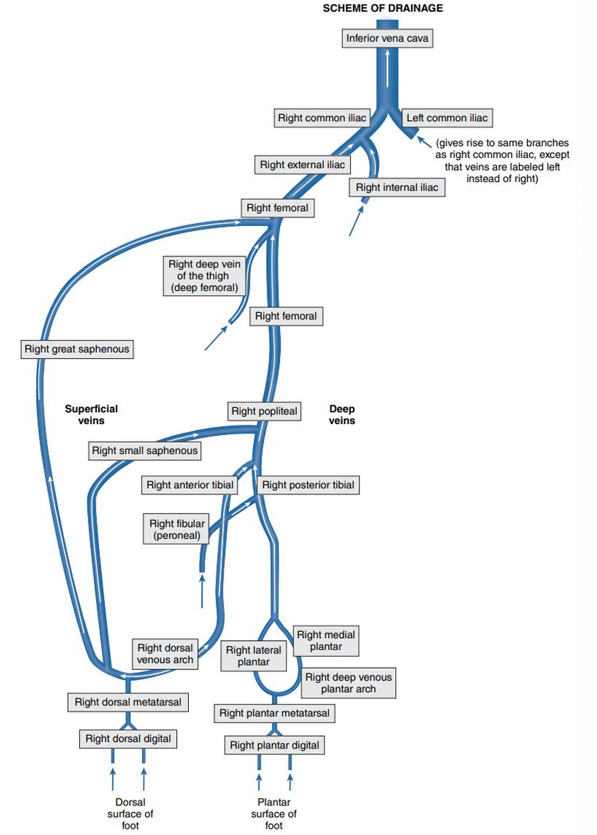
- Femoral V
- Accompany femoral A
- Extends from external iliac V at the region of the inguinal ligament 腹股沟韧带
- Femoral V → external iliac V → common iliac V → inferior vena cava → right atrium
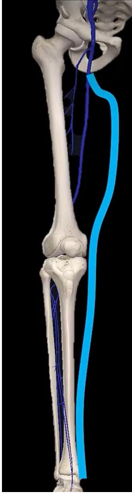
- Deep vein of thigh
- a tributary of femoral V 股深静脉
- Popliteal V
- continuation of femoral V
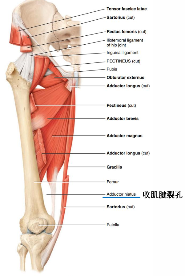
- Femoral V goes through adductor hiatus into popliteal V.
- 紫色Deep vein of thigh,浅蓝色Femoral V
- 图
- Veins of legs
- Anterior tibial V 胫前静脉
- Posterior Tibial V 胫后静脉
- Lies posterior to the medial malleolus in the ankle and ascends 内踝后侧上行
- Fibular V
- Lies posterior to the lateral malleolus in the ankle and ascends to empty into the posterior tibial V 外踝后侧上行
- Superficial veins
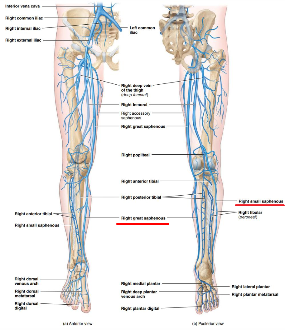
- Great saphenous veins
- Just deep to skin
- Begin at medial end of dorsal venous arch
- Pass anterior to medial malleolus of tibia 内踝前
- Ascends along medial side of leg and thigh
- Empty into femoral vein
- Have 10-20 valves, with more located in leg than thigh
- Varicose veins: enlarged, twisted veins
- Longest veins in body
- Small saphenous V
- Deep to skin
- Begin at lateral aspect of dorsal venous arch
- Ascend along posterior aspect of leg
- Empty into popliteal V, posterior to knee
- Have 9-12 valves
- 图
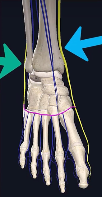
- 足背
- dorsal digital V → dorsal metatarsal V → dorsal venous arches
- 外侧Small saphenous V,Begin at lateral aspect of dorsal venous arch足背静脉弓
- 内侧Great saphenous V,Begin at medial end of dorsal venous arch
- 图
- Great saphenous veins
- 总结
- 图
- 图
- 分类
- 总结
- 图
- 图
- 基础
- 基础

All articles in this blog are licensed under CC BY-NC-SA 4.0 unless stating additionally.